
Page 297 - IJB-9-6
P. 297
International Journal of Bioprinting Progress in bioprinted ear reconstruction
Study Aim of study Study Animal Study focus 3D printing Components Printed Printed Cell nature/type Notable post- Assessment Findings Limitations and suggested
setting model (if technique shape material printing of success/ improvements
any) modifications integration
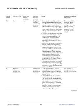
Mukherjee et al. To assess the degradation In vitro; Sheep Direct printing Extrusion Cells in bioink + Other shape PCL N/A None Histopathology; • Both porous 3D-printed PCL and hybrid The inclusion of GelMA-
(2021) [21] behavior and tissue in vivo (similar scaffold printed ultrasound scaffolds showed similar and homogenous HAMA with the PCL
compatibility of hybrid animal fascial together; scaffold scan; micro-CT degradation in vitro. In vivo, they exhibited demonstrated better
scaffolds (PCL-hydrogel) anatomy) only scan; electron minimal irritation or inflammation in tissue ingrowth. However,
compared to single microscopy surrounding tissue over a 6-month period its impact on cellular
material PCL scaffolds in in an immunocompetent animal model that differentiation can only
vitro and in vivo. The study closely resembles human soft tissue biology, be evaluated in future
wanted to understand although the host response varied between experiments embedded
the biological reaction animals. with cells.
to printed scaffolds • The hybrid scaffolds had a higher percentage
(independent of stem cells) mass loss than control scaffolds due to
in an immunocompetent the presence of degrading hydrogels that
host. contributed to a higher initial weight.
However, the degradation profile was
dominated by PCL in both hybrid and PCL-
only scaffolds.
• SEM showed that degradation occurred from
the outer surface inward for each strand.
• In vivo, the scaffolds were well tolerated for
the duration of the experiment, with serial
ultrasound and CT scans showing minimal
reaction in surrounding subcutaneous
tissue over 6 months. Ex vivo, the scaffolds
displayed localized hyperemia with peripheral
pallor and pseudo-capsule formation,
consistent with a localized inflammatory
response, indicating good biocompatible
properties with no macroscopic differences
between test and control samples.
• Control specimens, when 3D-reconstructed,
had less tissue integration compared to all
test samples in both sheep, regardless of PCL
configuration. This may be attributed to the
presence of the hydrogel.
Tang et al. To explore the use of In vitro; Mice Direct printing Fused deposition Scaffold printed Resembling PLA Rabbit ear XX Histopathology; • The successful induction of auricular Short-term study and
(2021) [15] 3D printing to fabricate in vivo modeling first and then pinna chondrocytes; mechanical chondrogenesis in vivo was demonstrated thus long-term ability to
bioactive artificial animal (extrusion) seeded with cells grafts were also testing; electron using a photosensitive GelMA hydrogel to withstand immune response
auricular cartilage using taken from mice microscopy allow chondrocytes to bind to a customized was not tested.
chondrocyte-laden GelMA auricular scaffold.
and PLA for auricle • A biologic auricle with a PLA material as
reconstruction. the inner core for support was constructed.
This not only provides mechanical support
for cartilage regeneration for morphological
maintenance in vitro and in vivo, but also
allows the ester bonds of PLA to be slowly
hydrolyzed, providing sufficient time for the
engineered cartilage to mature and acquire
mechanical properties while gradually
replacing the degrading PLA scaffold.
(Continued)
Volume 9 Issue 6 (2023) 289 https://doi.org/10.36922/ijb.0898