
Page 463 - IJB-9-6
P. 463
International Journal of Bioprinting 3D bioprinting of in vitro cartilage tissue model
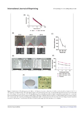
Figure 1. Optimization of 3D printing process for Alpha 1. (A) Dynamic viscosity vs. shear stress for Alpha 1, with and without medium (1:10). N = 3;
error bars show standard deviation. (B) Initial typical images of 3D-printed grid structure (30 × 30 mm) produced by different conical nozzle sizes (400
μm—22G, 250 μm—25G, and 200 μm—27G). (C) Quantification of the printed filament width of Alpha 1 using different nozzle sizes. N = 3; error bars
show standard deviation. (D) Typical images of printed Alpha 1 filaments at different extrusion pressures at constant printing speed of 10 mm/s using a
25G conical nozzle. (E) Correlation of printing speed with filament width at extrusion pressures of 8 and 10 kPa. (F) CAD design of printed cylindrical
structures (5 mm diameter, 1 mm thickness with 60% infill density) and images of printed structures in a 12-well plate.
Volume 9 Issue 6 (2023) 455 https://doi.org/10.36922/ijb.0899