
Page 466 - IJB-9-6
P. 466
International Journal of Bioprinting 3D bioprinting of in vitro cartilage tissue model
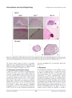
Figure 3. Top: PeptiInk Alpha 1 H&E staining. The top row shows lower-magnification images of cell-embedded hydrogels (scale bar: 200 µm); the arrows
on the lower-magnification images show cell cluster formations on the PeptiInk surface. The bottom row shows the magnified sections indicated by blue
boxes in the top row images (scale bar: 50 µm). Bottom: H&E staining of the 3D chondrocyte pellet over days 7 and 14 (scale bar: 50 µm).
The PeptiInk system showed low levels of intracellular somewhat upregulated but not statistically significantly
aggrecan on day 7, which then increased, especially at the (Figure 6C).
surface cell cluster formations (Figure 5). Negative controls
can be found in Figure S3 (Supplementary File). 4. Discussion
3.5. PCR analysis of chondrogenic markers The development of human in vitro models is crucial
In qRT-PCR, GADPH was used as housekeeping gene, to the further understanding of cartilage and diseases
and 3D pellet cultures were used as controls. Expression that affect it. Additionally, there is a need to move away
of specific cartilage markers, such as SOX9, COL2, and from animal models, which are not necessarily always
AGC, was assessed. Cells in control pellet cultures showed representative of human pathophysiology. Current
no significant changes in COL2, AGC, or SOX9 mRNA 3D-bioprinted models rely on the use of animal-derived
expression from day 7 to day 14 (Figure 6A). In contrast, materials such as gelatine or hyaluronan. These are natural
cells in Alpha 1 cultures showed an upregulation of all three bioinks that present multiple advantages such as enabling
mRNAs when compared to their corresponding control cell attachment and functionalization. However, they are
each time point (Figure 6B and C). Cells maintained animal-derived bioinks, which not only give rise ethical
in the Alpha 1 culture system exhibited a significant and sustainability issues in their production but also can
upregulation of COL2, AGC, and SOX9 mRNA expression interfere uncontrollably within the system. The use of
on day 7 (Figure 6B). On day 14, all mRNAs assessed were synthetic materials enables the possibility of creating a
Volume 9 Issue 6 (2023) 458 https://doi.org/10.36922/ijb.0899