Page 484 - IJB-9-6
P. 484

International Journal of Bioprinting                              Bioprinting cell-laden protein-based hydrogel


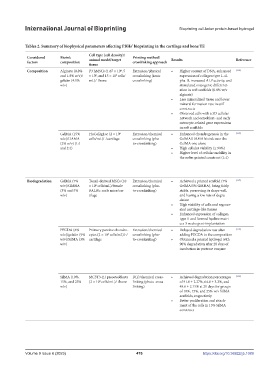
            Tables 2. Summary of biophysical parameters affecting PBHs’ bioprinting in the cartilage and bone TE
                                       Cell type (cell density)/
             Considered    Bioink      animal model/target   Printing method/  Results                Reference
             factors       composition                    crosslinking approach
                                       tissue
             Composition   Alginate (0.8%   P3 hMSCs (1.67 × 10 , 5   Extrusion/physical   •   Higher content of DNA, enhanced   [114]
                                                    6
                                                  6
                                          6
                           and 1.8% w/v)/  × 10 , and 15 × 10  cells/  crosslinking (ionic   expression of collagen type I–al-
                           gelatin (4.1%   mL)/-/bone     crosslinking)       pha-II, increased ALP activity, and
                           w/v)                                               stimulated osteogenic differenti-
                                                                              ation in soft scaffolds (0.8% w/v
                                                                              alginate)
                                                                          •   Less mineralized tissue and lower
                                                                              mineral formation rate in stiff
                                                                              constructs
                                                                          •   Observed cells with a 3D cellular
                                                                              network and osteoblast- and early
                                                                              osteocyte-related gene expressions
                                                                              in soft scaffolds
                           GelMA (15%   HuCol2gLuc (2 × 10   6  Extrusion/chemical   •   Enhanced chondrogenesis in the   [115]
                           w/v)/HAMA   cells/mL)/-/cartilage  crosslinking (pho-  GelMA/HAMA bioink over the
                           (2% w/v) (1:1                  to-crosslinking)    GelMA one alone
                           and 2:1)                                       •   High cellular viability (≥ 90%)
                                                                          •   Higher level of cellular mobility in
                                                                              the softer printed construct (1:1)


             Biodegradation  GelMA (7%   Tonsil-derived MSCs (10   Extrusion/chemical   •   Achieved a printed scaffold (7%   [117]
                           w/v)/GMHA   × 10  cells/mL)/female   crosslinking (pho-  GelMA/5% GMHA), being fairly
                                          6
                           (3% and 5%   BALB/c nude mice/car-  to-crosslinking)  stable, preserving its shape well,
                           w/v)        tilage                                 and having a low rate of degra-
                                                                              dation
                                                                          •   High viability of cells and regener-
                                                                              ated cartilage-like tissues
                                                                          •   Enhanced expression of collagen
                                                                              type II and formed hyaline matri-
                                                                              ces 3 weeks post-implantation
                           PEGDA (6%   Primary porcine chondro-  Extrusion/chemical   •   Delayed degradation rate after   [118]
                           w/v)/gelatin (9%  cytes (2 × 10  cells/mL)/-/  crosslinking (pho-  adding PEGDA to the composition
                                               6
                           w/v)/SilMA (3%  cartilage      to-crosslinking)  •   Obtained a printed hydrogel with
                           w/v)                                               90% degradation after 28 days of
                                                                              incubation in protease enzyme


                           SilMA (10%,   MC3T3-E1 preosteoblasts   DLP/chemical cross-  •   Achieved degradation percentages   [119]
                           15%, and 25%   (2 × 10  cells/mL)/-/bone  linking (photo-cross-  of 91.0 ± 2.27%, 64.8 ± 3.2%, and
                                            6
                           w/v)                           linking)            48.6 ± 2.15% at 21 days for groups
                                                                              of 10%, 15%, and 25% w/v SilMA
                                                                              scaffolds, respectively
                                                                          •   Better proliferation and attach-
                                                                              ment of the cells in 15% SilMA
                                                                              construct


            Volume 9 Issue 6 (2023)                        476                          https://doi.org/10.36922/ijb.1089
   479   480   481   482   483   484   485   486   487   488   489