
Page 485 - IJB-9-6
P. 485
International Journal of Bioprinting Bioprinting cell-laden protein-based hydrogel
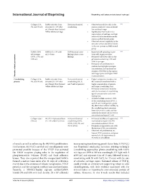
Collagen (1%, Rabbit articular chon- Extrusion/chemical • Observed mostly live cells in the [121]
3%, and 5% wt) drocytes (1 × 10 cells/ crosslinking porous constructs’ cross-sectional
6
mL)/female New Zealand live and dead image
White rabbits/cartilage • Significantly improved in vivo
regeneration of cartilage, and high
levels of GAGs distribution in the
porous scaffold-treated group
• Observed severe irregularity in the
articular surface and defect lesions
in the non-porous scaffold-treated
group
GelMA (20% hMSCs (2 × 10 cells/ DLP/chemical cross- • Improved cell spreading in por- [125]
6
wt)/porogen mL)/-/bone linking (photo-cross- • tions with larger pore sizes
Porosity-related parameters • 3.0% wt porogen
(0.5%, 1.5%, and
linking)
Promoted cell cluster sizes in the
3.0% wt)
gel regions containing 1.5% and
Enhanced cell proliferation in
portions having higher porogen
concentration, and increased ex-
pression of RUNX2 in the regions
2 concentrations
Crosslinking Collagen (1%, Rabbit articular chon- Extrusion/chemical • with larger pores and higher BMP- [121]
Higher compressive modulus for
process 3%, and 5% wt) drocytes (1 × 10 cells/ crosslinking (0.1, 1, the construct crosslinked with a
6
mL)/female New Zealand and 5 mM of genipin) higher concentration of genipin
White rabbits/cartilage and longer crosslinking time
• Enhanced compressive modulus
with the increment of crosslinking
agent’s concentration and cross-
linking time
• Achieved cellular survival > 90%
at the crosslinking times of 0.5, 1,
and 6 h at 0.1 mM genipin, gradu-
ally diminished cellular viability by
the crosslinking time’s extension
from 0.5 to 6 h at 1 mM, and dra-
matically decreased cell survival at
concentrations beyond 5 mM
Collagen type I MC3T3-E1 preosteoblasts Extrusion/chemical • Enhanced stability and augmented [133]
(5% wt) (5 × 10 cells/mL)/-/bone crosslinking (0.1%, mechanical strength in the TA
6
0.25%, 0.5%, 1%, and concentration above 0.5% wt
3% wt of TA) • Diminished degradation rate with
the increase of TA concentration
• Observed cell proliferation, en-
hanced DNA expression, and great
survival of cells (approximately
95–96%) in all scaffolds
of vessels, as well as enhancing the HUVECs proliferation. incorporating transforming growth factor beta-3 (TGF-β )
3
Furthermore, HUVEC’s survival and vasculogenesis were was bioprinted employing extrusion printing technique
promoted possibly because of the VEGF that activated (crosslinking approaches: photo-crosslinking with 0.05%
the VEGF receptors playing roles in the regulation of w/v Irgacure 2959 at 365 nm wavelength, UV light with 5
phosphoinositide 3-kinase (P13K) and focal adhesion mW/cm , and exposure time of 15 min, followed by ionic
2
kinase. They also reported that the encapsulated hMSCs crosslinking with 50 mM CaCl for 15 min) with the aim
2
formed a mature bone niche after 21 days of cultivation of articular cartilage repair. Sustained release of TGF-β
3
mainly due to the existence of silicate nanoplatelets, VEGF could provide an environment supporting robust in vitro
presence, and the osteogenic medium’ sustained perfusion. chondrogenesis, with little evidence associated with the
Recently, a hBMSCs-loaded alginate sulfate/GelMA mineralization or hypertrophy over extended periods of
bioink (1% and 10% w/v) (cell density: 20 × 10 cells/mL) culture. Moreover, in vivo 4-week results that included
6
Volume 9 Issue 6 (2023) 477 https://doi.org/10.36922/ijb.1089