
Page 164 - v11i4
P. 164
International Journal of Bioprinting 3D-printed microstructure for bacteriostasis
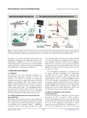
Figure 1. Schematic showing the fabrication of finely tuned bacteriostatic surfaces to inhibit bacterial growth. Bionic microstructures were designed to
simulate shark skin using SolidWorks 2019. A photosensitive resin, IP-S, was attached to the substrate surface after pretreatment by oxygen plasma and
two-photon polymerization. Finally, the designed bionic bacteriostatic microstructures were accurately constructed on the substrate surface.
technology can be used as a powerful tool to explore the (3 or 5 μm) and width (2 or 5 μm), yet with different lengths
underlying mechanisms and influencing factors of the (4, 8, 12, and 16 μm), were arranged in parallel with a 2- or
bacteriostatic capabilities of various microstructures, 5-μm-wide gap. Holes with a depth of 3 μm and different
thereby deepening the understanding of the bacteriostatic diameters (2, 3, and 4 μm) were arranged in parallel, with
mechanisms of micro- and nano-structures and broadening the spacing between two adjacent holes equal to the hole
their applications in various aspects of daily life. diameter. The centers of every three adjacent holes formed
an equilateral triangle.
2. Materials and methods
For microstructure fabrication, a two-photon laser
2.1. Materials 3D printer (Photonic Professional GT2, Nanoscribe,
Photo-sensitive resin (IP-S) and glass substrates were Germany) and IP-S were employed. Initially, glass
obtained from Nanoscribe, Germany. S. mutans was substrates were pre-treated by oxygen plasma (PDC-001,
provided by the School & Hospital of Stomatology, Harrick Plasma, USA) to enhance the affinity between
Wuhan University, China. Brain-heart infusion (BHI) was IP-S and the substrates. Subsequently, IP-S was dropped
obtained from Phygene, China. The reagents of crystal on the glass substrates and placed into the 3D printer for
violet and glutaraldehyde were purchased from Sigma- constructing specific microstructures according to design.
Aldrich, USA. Fluorescent SYTO 9 reagent, phosphate- Finally, PGMEA and isopropanol were employed to
buffered saline (PBS, 1×, pH = 7.2), and anhydrous ethanol develop those microstructures, and PBS was used to wash
were obtained from Thermo-Fisher Scientific, USA. Agar, the substrates to remove excess organic reagents.
propylene glycol monomethyl ether acetone (PGMEA),
and isopropanol were purchased from Aladdin, China. 2.3. Bacteria cultivation on the surfaces
of microstructures
2.2. Design and fabrication of microstructures on S. mutans was cultured in a BHI solution growth medium
glass substrates and grown at 37°C overnight in static conditions.
The design of the biomimetic microstructures (shark skin Substrates with different microstructures were placed into
denticle simulation and corresponding simplified indented six-well plates and sterilized with ultraviolet (UV) light
micro-strips) or circular holes microstructure was prepared for 30 min before experiments. Subsequently, S. mutans
by SolidWorks 2019 (Dassault Systems – SolidWorks suspension made with BHI broth concentration of 1 ×
Corporation, USA). Various strips with the same height 10 /mL was inoculated into each well of the six-well
6
Volume X Issue X (2025) 156 doi: 10.36922/IJB025150135