
Page 165 - v11i4
P. 165
International Journal of Bioprinting 3D-printed microstructure for bacteriostasis
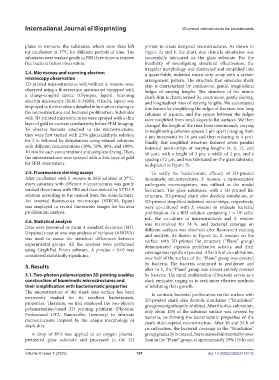
plates to immerse the substrates, which were then left printer to create designed microstructures. As shown in
for incubation at 37°C for different periods of time. The Figure 2a and b, the shark skin denticle simulation was
substrates were washed gently in PBS three times to remove successfully fabricated on the glass substrate. For the
free bacteria before observation. feasibility of investigating structural effectiveness, the
irregular morphology was abstracted and simplified into
2.4. Microscopy and scanning electron a quantifiable, indented micro-strip array with a certain
microscopy observation arrangement pattern. The structure that simulates shark
3D-printed microstructures with/without S. mutans were skin is characterized by continuous, gentle, longitudinal
observed using a fluorescence microscope equipped with bulges of varying lengths. The structure of the mimic
a charge-coupled device (Olympus, Japan). Scanning shark skin is characterized by continuous, gently sloping,
electron microscopy (SEM; S-3400N, Hitachi, Japan) was and longitudinal rises of varying lengths. We accentuated
employed to further obtain detailed information relating to this feature by simplifying the ridges of the rises into long
the microstructures and bacterial proliferation. Substrates columns of squares, and the spaces between the ridges
with 3D-printed microstructures were sprayed with a thin were simplified from small slopes to flat surfaces. We then
layer of gold to increase conductivity before SEM imaging. changed the length of the rises from continuously varying
To observe bacteria attached to the microstructures, to neighboring columns spaced 2 µm apart (ranging from
they were first treated with 2.5% glutaraldehyde solution 4 µm increments to 16 µm and then reducing to 4 µm).
for 2 h, followed by dehydration using ethanol solutions Finally, this simplified structure featured seven parallel
with different concentrations (30%, 50%, 80%, and 100%, indented micro-strips of varying lengths (4, 8, 12, and
10 min for each concentration) and complete drying. Then, 16 µm), with a height of 3 µm, a width of 2 µm, and a
the microstructures were sprayed with a thin layer of gold spacing of 2 µm, and was fabricated on the glass substrate,
for SEM observation. as depicted in Figure 2b.
2.5. Fluorescence staining assays To verify the bacteriostatic efficacy of 3D-printed
After incubation with S. mutans in BHI solution at 37°C, biomimetic microstructures, S. mutans, a representative
every substrate with different microstructures was gently pathogenic microorganism, was utilized as the model
washed three times with PBS and then stained by SYTO 9 bacterium. The glass substrates, with a 3D-printed flat
solution according to the manual from the manufacturer. structure, 3D-printed shark skin denticle simulation, or
An inverted fluorescence microscope (NIKON, Japan) 3D-printed simplified indented micro-strips, respectively,
was employed to record fluorescent images for bacteria were co-cultured with S. mutans to evaluate bacterial
proliferation analysis. proliferation. In a BHI solution containing 1 × 10 cells/
6
mL, the co-culture of microstructures and S. mutans
2.6. Statistical analysis was maintained for 24 h, and bacterial coverage on
Data were presented as mean ± standard deviation (SD). different surfaces was observed after fluorescent staining
Unpaired t-test or one-way analysis of variance (ANOVA) and analysis. As shown in Figure 2c, S. mutans on the
was used to assess the statistical differences between surface with 3D-printed flat structure (“Plane” group)
experimental groups. All the analyses were performed demonstrated vigorous proliferative activity, and their
using GraphPad Prism software. A p-value < 0.05 was coverage was rapidly expanded. After 8 h of co-cultivation,
considered statistically significant.
over half of the surface of the “Plane” group was covered
3. Results by bacteria. The bacteria continued to proliferate and
after 16 h, the “Plane” group was almost entirely covered
3.1. Two-photon polymerization 3D printing enables by bacteria. The rapid proliferation of bacteria serves as a
construction of biomimetic microstructures and stark reminder, urging us to seek more effective methods
their simplification with bacteriostatic properties of inhibiting their growth.
The microstructure of the shark skin surface has been In contrast, bacterial proliferation on the surface with
extensively studied for its excellent bacteriostatic 3D-printed shark skin denticle simulation (“Simulation”
properties. Therefore, we first employed the two-photon group) was significantly inhibited. After 8 h of co-cultivation,
polymerization-based 3D printing platform (Photonic only about 10% of the substrate surface was covered by
Professional GT2, Nanoscribe, Germany) to fabricate bacteria, confirming the bacteriostatic properties of the
microstructures inspired by the unique morphology of shark skin-inspired microstructure. After 16 and 24 h of
shark skin. co-cultivation, the bacterial coverage in the “Simulation”
A drop of IP-S was applied to an oxygen plasma- group gradually increased, but remained substantially lower
pretreated glass substrate and processed in the 3D than in the “Plane” group, at approximately 25% (16 h) and
Volume X Issue X (2025) 157 doi: 10.36922/IJB025150135