
Page 253 - v11i4
P. 253
International Journal of Bioprinting Deep learning-based 3D digital model of fetal heart
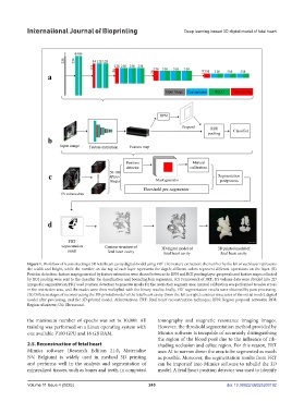
Figure 1. Workflow of reconstructing a 3D fetal heart cavity digital model using FRT. (A) Feature extraction: the number by the left of each layer represents
the width and height, while the number on the top of each layer represents the depth; different colors represent different operations on the layer. (B)
Position detection: feature maps generated by feature extraction were shared between the RPN and ROI pooling layer; proposals and feature maps collected
by ROI pooling were sent to the classifier for classification and bounding box regression. (C) Framework of FRT: US volume data were divided into 2D
images for segmentation; FRT used position detection to generate masks for the restriction segment area; manual calibration was performed to solve errors
in the restriction area, and the masks were then multiplied with the binary results; finally, FRT segmentation results were obtained by post-processing.
(D) Different stages of reconstructing the 3D-printed model of the fetal heart cavity (from the left to right): contour structures of the initial model, digital
model after processing, and the 3D-printed model. Abbreviations: FRT: Fetal heart reconstruction technique; RPN: Region proposal networks; ROI:
Region of interest; US: Ultrasound.
the maximum number of epochs was set to 10,000. All tomography and magnetic resonance imaging images.
training was performed on a Linux operating system with However, the threshold segmentation method provided by
one available P100 GPU and 16 GB RAM. Mimics software is incapable of accurately distinguishing
the region of the blood pool due to the influence of rib-
2.5. Reconstruction of fetal heart shading occlusion and celiac region. For this reason, FRT
Mimics software (Research Edition 21.0, Materialise uses AI to narrow down the area to be segmented as much
NV, Belgium) is widely used in medical 3D printing as possible. Moreover, the segmentation results from FRT
and performs well in the analysis and segmentation of can be imported into Mimics software to rebuild the 3D
mineralized tissues, such as bones and teeth, in computed model. A fetal heart position detector was used to identify
Volume 11 Issue 4 (2025) 245 doi: 10.36922/IJB025200192