
Page 255 - v11i4
P. 255
International Journal of Bioprinting Deep learning-based 3D digital model of fetal heart
Since the assessment of performance on 2D images 3. Results
alone is insufficient to verify the accuracy of spatial 3.1. Segmentation performance
structures of the 3D digital model, digital models rebuilt For reconstructing the 3D digital model of the fetal heart,
by different methods (e.g., manual reconstruction by identifying the blood pool in fetal hearts was the most
doctors of varying experience and deep learning-based important step in the process. Accuracy, interpretability,
methods) were printed into physical models. The long and variability were all important indicators for
diameter of the left ventricle (LDLV), long diameter reconstruction. To investigate the accuracy of FRT, the
of the right ventricle (LDRV), long diameter of the left mIOU/DSC of segmentations from three different views—
atrium (LDLA), the transverse diameter of the left atrium using the thresholding method in Mimics software—were
(TDLA), long diameter of the right atrium (LDRA), 0.242/0.381 for FCV, 0.165/0.278 for OTV, and 0.121/0.214
and transverse diameter of right atrium (TDRA) were for TVV, respectively. Using the FRT method, there was
measured based on the physical model using a vernier a significant improvement in the performance from
caliper (Figure 6F). In addition, the same measurements different views (FCV: 0.701/0.823; OTV: 0.681/0.809;
were performed by sonographers of varying experience TVV: 0.602/0.746). Furthermore, smoothing and removal
(junior doctor: <3 years of experience; middle doctor: 3–6 of free small objects also enhanced performance based
years of experience; senior doctor: >6 years of experience) on FCV and OTV (FCV: 0.714/0.832; OTV: 0.687/0.813)
on US volume data using the Voluson E10 US system. after calibration. In summary, FRT performance is better
Notably, amplifying fine structures is an advantage of the based on OTV and TVV and worse based on FCV relative
3D-printed model. Direct comparison of specific values is to segmentation conducted by junior doctors. Details of
not scientific, as the length of each part changes with the the evaluation indicators of the segmentation results using
magnification. Thus, with LDLV as the benchmark, the different methods are displayed in Table 2.
rest of the metrics were converted into ratios relative to 3.2. Interpretability of results
LDLV. Finally, a one-way analysis of variance (ANOVA) To further investigate the interpretability of the significant
was performed for comparisons among clinical evaluation improvements achieved by the position detector, we
values in proportion. hypothesized that it reduces the influences of noise,
The performance of the segmentation task was shadow, and other factors in US images by limiting the area
evaluated using Python data analysis and manipulation of image processing. The grayscale distribution maps of the
tools, such as Numpy, Pandas, and Matplotlib. For segmentation area indicated that there was a significant
evaluation of the performance of the 3D structure, one-way decrease in the number of pixels in the grayscale range of
ANOVA was performed for comparisons among multiple 20–40 (Figures 2 and 3).
groups using GraphPad PRISM 5.0. For all experiments, Combined with the original images, these reduced
significance was defined as: *p < 0.05, **p < 0.01, and pixels were mainly distributed in the shadow and
***p < 0.001. No statistical method was used to noise of the non-target region. The recall rate of FRT
predetermine the sample size. significantly improved, and the positive prediction rate
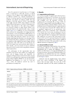
Table 2. Segmentation performance of different methods
Method FCV OTV TVV
IOU DSC IOU DSC IOU DSC
Threshold 0.242 0.381 0.165 0.278 0.121 0.214
U-Net 0.712 0.830 0.593 0.740 0.431 0.591
Junior doctor 0.798 0.887 0.658 0.789 0.530 0.684
FRT-Default 0.701 0.823 0.681 0.809 0.602 0.746
FRT-IA 0.714 0.832 0.687 0.813 0.599 0.744
Note: IOU and DSC were used to evaluate the segmentation performance across different methods (rows); “FRT-Default” refers to segmentation by
FRT using default parameters without manual interaction; “FRT-IA” refers to segmentation by FRT with manual interaction. Abbreviations: DSC: Dice
similarity coefficient; FCV: Four-chamber view; FRT: Fetal heart reconstruction technique; IOU: Intersection over union; OTV: Outflow tract view;
TVV: Three-vessel view.
Volume 11 Issue 4 (2025) 247 doi: 10.36922/IJB025200192