
Page 260 - v11i4
P. 260
International Journal of Bioprinting Deep learning-based 3D digital model of fetal heart
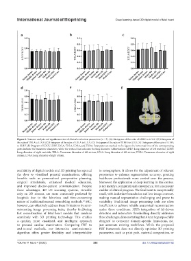
Figure 6. Variance analysis and significance test of clinical evaluation parameters (n = 3). (A) Histogram of the ratio of LDRV to LDLV. (B) Histogram of
the ratio of TDLA to LDLV. (C) Histogram of the ratio of LDLA to LDLV. (D) Histogram of the ratio of TDRA to LDLV. (E) Histogram of the ratio of LDRA
to LDLV. (F) Diagram of LDLV, LDRV, LDLA, TDLA, LDRA, and TDRA. Four parts are marked in the figure: the horizontal lines of the corresponding
parts indicate the transverse diameters, while the vertical line indicates the long diameter. Abbreviations: LDLV: Long diameter of left ventricle; LDRV:
Long diameter of right ventricle; TDLA: Transverse diameter of left atrium; LDLA: Long diameter of left atrium; TDRA: Transverse diameter of right
atrium; LDRA: Long diameter of right atrium.
availability of digital models and 3D printing has opened to sonographers. It allows for the adjustment of relevant
the door to visualized prenatal examinations, offering parameters to enhance segmentation accuracy, granting
benefits such as personalized preoperative planning, healthcare professionals more control over the process.
surgical simulations, enhanced medical education, Moreover, the application of deep learning in this context
and improved doctor–patient communication. Despite is not merely a computational convenience, but a necessary
these advantages, 4D US scanning systems, viewable enabler of clinical progress. The fetal heart is exceptionally
only on 2D screens, are more commonly preferred by small, with indistinct boundaries and low image contrast,
hospitals due to the laborious and time-consuming making manual segmentation challenging and prone to
nature of traditional manual remodeling methods. FRT, variability. Traditional image processing tools are often
28
however, can effectively address these limitations by semi- insufficient to achieve reliable anatomical reconstruction
automating image processing tasks, thereby facilitating under these conditions. FRT’s integration of AI-driven
fast reconstruction of fetal heart models that combine detection and interactive thresholding directly addresses
seamlessly with 3D printing technology. This enables these challenges, demonstrating that AI can be purposefully
a quicker, more visualized, and intelligent approach designed to overcome domain-specific limitations, not
to prenatal cardiac examination. Comparing FRT to just automate existing workflows. While the proposed
end-to-end methods, our interactive semi-automatic FRT framework does not directly optimize 3D printing
algorithm offers greater flexibility and interpretability parameters, such as print path, material composition, or
Volume 11 Issue 4 (2025) 252 doi: 10.36922/IJB025200192