
Page 256 - v11i4
P. 256
International Journal of Bioprinting Deep learning-based 3D digital model of fetal heart
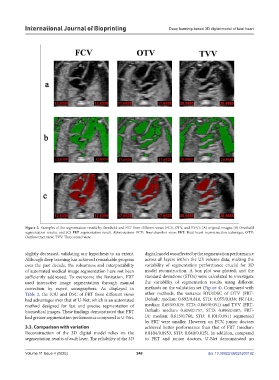
Figure 2. Examples of the segmentation results by threshold and FRT from different views (FCV, OTV, and TVV): (A) original images; (B) threshold
segmentation results, and (C) FRT segmentation result. Abbreviations: FCV: Four-chamber view; FRT: Fetal heart reconstruction technique; OTV:
Outflow tract view; TVV: Three-vessel view.
slightly decreased, validating our hypothesis to an extent. digital model was affected by the segmentation performance
Although deep learning has achieved remarkable progress across all layers within the US volume data, making the
over the past decade, the robustness and interpretability variability of segmentation performance crucial for 3D
of automated medical image segmentation have not been model reconstruction. A box plot was plotted, and the
sufficiently addressed. To overcome the limitation, FRT standard deviations (STDs) were calculated to investigate
used interactive image segmentation through manual the variability of segmentation results using different
correction by expert sonographers. As displayed in methods on the validation set (Figure 4). Compared with
Table 2, the IOU and DSC of FRT from different views other methods, the variance IOU/DSC of OTV (FRT-
had advantages over that of U-Net, which is an automated Default: median: 0.692/0.818, STD: 0.055/0.038; FRT-IA:
method designed for fast and precise segmentation of median: 0.693/0.819, STD: 0.069/0.051) and TVV (FRT-
biomedical images. These findings demonstrated that FRT Default: median: 0.609/0.757, STD: 0.096/0.087; FRT-
had greater segmentation performance compared to U-Net. IA: median: 0.613/0.760, STD: 0.101/0.091) segmented
by FRT were smaller. However, on FCV, junior doctors
3.3. Comparison with variation achieved better performance than that of FRT (median:
Reconstruction of the 3D digital model relies on the 0.8104/0.8953, STD: 0.040/0.025). In addition, compared
segmentation results of each layer. The reliability of the 3D to FRT and junior doctors, U-Net demonstrated no
Volume 11 Issue 4 (2025) 248 doi: 10.36922/IJB025200192