
Page 259 - v11i4
P. 259
International Journal of Bioprinting Deep learning-based 3D digital model of fetal heart
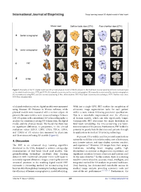
Figure 5. Examples of the 3D digital model and 3D-printed physical model of the blood pool in the fetal heart reconstructed by different methods based
on the whole heart structure, OTV, and FCV: (A) manually reconstructed by a senior sonographer, (B) manually reconstructed by a junior sonographer,
(C) reconstructed using FRT, and (D) reconstructed using U-Net. Abbreviations: FRT: Fetal heart reconstruction technique; OTV: Outflow tract view;
FCV: Four-chamber view.
of clinical evaluation values, digital models were measured With just a single GPU, FRT enables the completion of
using Measure 3D Distance in Mimics software, while ultrasonic image segmentation tasks for each patient
physical models were measured with a vernier caliper. At within a mere minute following parameter specification.
present, the same metrics were measured using a Voluson This is a remarkable improvement over the efficiency
E10 US system with conventional 2D echocardiography to of human experts, which can take significantly longer.
analyze the consistency among US volume data, the digital Consequently, FRT overcomes two major limitations in
model, and the physical model. We found that there was fetal heart remodeling: the time-consuming and labor-
no significant difference, in proportion, in the clinical intensive nature of the process. Furthermore, FRT holds the
evaluation values (LDLV, LDRV, LDLA, TDLA, LDRA, potential to greatly benefit clinicians and patients through
and TDRA) of US volume data measured by physicians its applications in medical 3D printing technology.
and those measured using 3D models (Figure 6).
At present, US is widely used for prenatal examinations
4. Discussion across the world due to its relative safety, cost-effectiveness,
non-invasive nature, real-time display, operator comfort,
The FRT is an advanced deep learning algorithm and experience. However, US images have their unique
22
developed in the USA, designed to achieve cutting-edge limitations, including lower imaging quality, high
reconstruction of fetal heart blood pool models. This dependence on operator or diagnostician experience, and
groundbreaking technology combines deep learning significant variations between observers and systems used
detectors with traditional computer vision techniques to by the same observer. In this context, there is a pressing
23
accurately segment ultrasonic images, ensuring the utmost need for a more objective, accurate, visual, intelligent, and
structural precision in the resulting digital model. FRT integrated method for US analysis in prenatal healthcare.
represents a pioneering method for reconstructing fetal Deep learning has demonstrated remarkable success in
heart models through deep learning technology, surpassing medical image analysis, providing automatic tools and
the efficiency of human sonographers in model rebuilding. state-of-the-art performance. 15,17,18,24–27 Furthermore, the
Volume 11 Issue 4 (2025) 251 doi: 10.36922/IJB025200192