
Page 257 - v11i4
P. 257
International Journal of Bioprinting Deep learning-based 3D digital model of fetal heart
significant advantage across the three views (FCV: 3D digital model and 3D-printed physical model
median: 0.6721/0.804, STD: 0.076/0.055; OTV: median: (Figure 5). Our findings indicate that the physical
0.654/0.791, STD: 0.142/0.138; TVV: median: 0.547/0.707, model can intuitively display the spatial structures and
STD: 0.158/0.165). These results indicated that FRT had morphologic characteristics of the blood pool in the fetal
achieved a non-inferior performance compared with heart. Likewise, the 3D-printed physical models were
junior doctors, consistent with the results in Table 2. well-matched with digital models, and the FRT model
3.4. Analysis of spatial structures of 3D models achieved the best accuracy among these models based on
All cases in the validation set were successfully rebuilt the structure of the whole heart, as well as the OTV and
and printed using FRT to assess the accuracy of the FCV of the fetal heart. To further investigate the accuracy
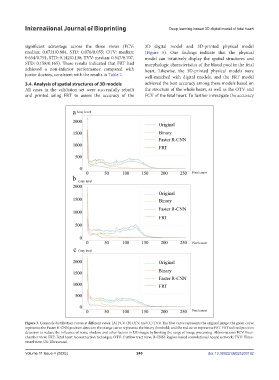
Figure 3. Grayscale distribution curves at different views: (A) FCV, (B) OTV, and (C) TVV. The blue curve represents the original image; the green curve
represents the Faster-R-CNN position detector; the orange curve represents the binary threshold; and the red curve represents FRT. FRT utilized position
detection to reduce the influence of noise, shadow, and other factors in US images by limiting the range of image processing. Abbreviations: FCV: Four-
chamber view; FRT: Fetal heart reconstruction technique; OTV: Outflow tract view; R-CNN: Region-based convolutional neural network; TVV: Three-
vessel view; US: Ultrasound.
Volume 11 Issue 4 (2025) 249 doi: 10.36922/IJB025200192