
Page 402 - v11i4
P. 402
International Journal of Bioprinting Bioprinted liver dECM/GelMA tumor model
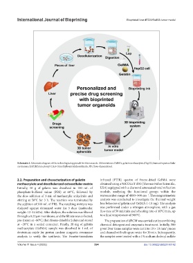
Schematic 1. Schematic diagram of the technological approach for this research. Abbreviations: GelMA, gelatin methacrylate; HepG2, human hepatocellular
carcinoma; LAP, lithium phenyl-2,4,6-trimethylbenzoylphosphinate; 3D, three-dimensional.
2.2. Preparation and characterization of gelatin infrared (FTIR) spectra of freeze-dried GelMA were
methacrylate and decellularized extracellular matrix obtained using a NICOLET iS50 (Thermo Fisher Scientific,
Initially, 10 g of gelatin was dissolved in 100 mL of USA) equipped with a diamond attenuated total reflection
phosphate-buffered saline (PBS) at 60°C, followed by module, analyzing the functional groups within the
–1
the slow addition of 3 mL of methacrylic anhydride and wavenumber range of 4000–500 cm . Thermogravimetric
stirring at 50°C for 3 h. The reaction was terminated by analysis was conducted to investigate the thermal weight
the addition of 500 mL of PBS. The resulting mixture was loss behavior of gelatin and GelMA (~10 mg). The analysis
dialyzed against deionized water for 3 days (molecular was performed under a nitrogen atmosphere, with a gas
weight: 12–14 kDa). After dialysis, the solution was filtered flow rate of 50 mL/min and a heating rate of 10°C/min, up
through a 0.22 µm membrane, and the filtrate was collected, to a final temperature of 500°C.
pre-frozen at –80°C, then freeze-dried for 2 days and stored The preparation of dECM was carried out by combining
at –20°C in a sealed container. Finally, 20 mg of gelatin chemical detergent and enzymatic treatment. Initially, 500
methacrylate (GelMA) sample was dissolved in 1 mL of g wet liver tissue samples were cut into 10 × 10 mm pieces
2
deuterium oxide for proton nuclear magnetic resonance and cleaned with ultrapure water for 30 min. Subsequently,
analysis to verify the synthesis. The Fourier-transform the samples were treated with a 1% sodium dodecyl sulfate
Volume 11 Issue 4 (2025) 394 doi: 10.36922/IJB025160142