
Page 407 - v11i4
P. 407
International Journal of Bioprinting Bioprinted liver dECM/GelMA tumor model
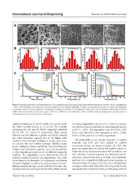
Figure 2. Hydrogel preparation and characterization. (A) Scanning electron microscopy images of five different hydrogels. Scale bar: 100 μm; magnification
= 100×. (B) Distribution of macroporous structure diameter of five different hydrogels. (C) Shear rate-dependent viscosity, (D) shear rate-dependent
temperature, and (E) viscosity-dependent temperature of different biomaterial ink formulations. Notes: GM: 10% (w/v) gelatin methacrylate (GelMA);
GM/G: 10% (w/v) GelMA and 5% (w/v) gelatin; GM/G/d-1, GM/G/d-3, and GM/G/d-5: GM/G combined with decellularized extracellular matrix at
concentrations of 1%, 3%, and 5% (w/v), respectively.
scaffold exhibited an Sr of 8.92 within this period, while the slowest degradation rate at 14.52 ± 1.03%. In contrast,
the GM/G scaffold showed an Sr of 8.05. The scaffolds the GM/G/d-5 group exhibited the fastest degradation rate
containing 1%, 3%, and 5% dECM composites exhibited at 39.74 ± 4.03%. The degradation rates for GM/G, GM/
Srs of 7.49, 7.01, and 6.13, respectively. These results G/d-1, and GM/G/d-3 were measured at 19.9 ± 2.39%,
indicate that the addition of gelatin and dECM reduced 23.85 ± 2.67%, and 32.14 ± 3.01%, respectively.
the water absorption capacity due to the higher solid
content in the hydrogel, resulting in smaller pore sizes. Biocompatibility is an essential property of scaffold
In contrast, the pure GelMA hydrogel exhibited larger materials, and L929 cells were applied for scaffold
pores, leading to a faster equilibrium rate of swelling and cytotoxicity testing. As shown in Figure 3E, L929 cells
a higher water absorption capacity. Figure 3D presents the grew and proliferated normally in the scaffold extract, in
in vitro degradation properties of the different composite which the cell survival rate was above 90% at both 24- and
scaffolds. The degradation curves demonstrate that all 48-h intervals. Consistent growth was observed whether
scaffolds exhibited relatively fast degradation. This could cells were cultured in the extract or fresh culture medium,
be attributed to the disruption of the physical cross-linking and there was no significant difference between groups. As
structure of gelatin, as well as the reduced degree of cross- shown in Figure S4, Supporting Information, the live/dead
linking caused by the high content of dECM. Over the 9-day staining of cells at 48 h where the L929 cell growth density
experimental observation period, the GM scaffold showed was similar within the field of view, and the group with 5%
Volume 11 Issue 4 (2025) 399 doi: 10.36922/IJB025160142