Page 63 - IMO-2-1
P. 63

Innovative Medicines & Omics                                         Flavonoids against glycosidic hydrolase


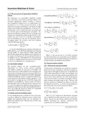
            2.3.2. The assessment of α-glucosidase inhibition                     1   K      I     1  1
            activity                                           Competitiveinhibition: =  m   1+     +   (II)

                                                                                                  S
                                                                                  v V max    K i     V
            The assessment of  α-glucosidase inhibition activity                                       maax
            was conducted as described in previous studies, with
            necessary adjustments made.  Initially, a mixture                       1   K m  1    1    [ ] I 
                                      14
                                                                                                     
            was prepared by adding 50  μL of  α-glucosidase at a   Uncompetitive inhibition:   =  V  [ ] S  + V max     1+ K i   
                                                                                    v
            concentration of 2 U/mL, followed by 50  μL of the                           max
            inhibitor at varying concentrations, and 80 μL of buffer     Noncompetitive inhibition:       (III)
            solution. This mixture was incubated for 10 min at 37°C.
                                                                           I
                                                                                           I
            Subsequently, 50 μL of pNPG (20 mM) was added, and   1 =  K m    1+      1  +  1 1    1+      (IV)
            the contents were mixed thoroughly and shaken. The   v V max    K   S   V max    K
                                                                            i
                                                                                            i
            reaction was allowed to continue at 37°C for 20 min, after
            which it was concluded by adding 100 μL of Na CO . The
                                                                                         I
                                                                                                         I
                                                      3
                                                  2
                                                                              1
            solution was then diluted to a suitable concentration,   Mixedinhibition: =  K m    1+      1  +  1    1+

                                                                                             S
            and the absorbance at 540  nm was measured with a                 v V max    K i     V max    K is
            microplate reader. The inhibition rate was calculated                                         (V)
            using the following equation:                             K      I
                             (A -A )                           Slope=  m   1+                         (VI)
            Inhibitionrate%=1-[  3  4  ] 100            (I)          V max    K
                                     ×
                                                                              i
                             (A -A )
                                  2
                               1
              A  served as the blank group, consisting of enzyme and     1
                                                                                I
                1
            buffer, while A  acted as the blank control group, containing   Y intercept = V    1+ K      (VII)
                       2
            only  the buffer.  A  represented  the sample group that    max    is
                           3
            included the enzyme and the inhibitor, and A  functions as
                                                4
            the sample control group, which was made up of buffer and   Where v is the reaction rate; K , K, and K  represent
                                                                                           m
                                                                                                     is
                                                                                              i
            the inhibitor. The IC  value indicated the concentration of   the Michaelis-Menten, free and bound enzyme inhibition
                            50
            the inhibitor needed to reduce the enzyme activity by half.   constants, respectively; [S] stands for substrate concentration;
            Acarbose was established as a control substance.   [I] represents the concentration of the inhibitor.
            2.4. Correlation analysis                          2.6. Characterization method
            The research focused on the concentration-effect   2.6.1. Fluorescence spectrum analysis
            relationships  of  various  solvent  extracts  regarding   A fluorescence quenching test was conducted to study the
            their ability to inhibit the functions of  α-amylase and   interaction between flavonoids in FBSJ and α-amylase or
            α-glucosidase. Grey relation analysis (GRA) was performed   α-glucosidase, with slightly modifications.  α-amylase (0.58
                                                                                               17
            using Statistical Package for the Social Sciences (SPSS) Pro,   U/mL) and α-glucosidase (2 U/mL) were incubated with
            utilizing the peak areas obtained from the HPLC fingerprint   different concentrations of quercetin/kaempferol in the water
            profiles of the different extracts, in conjunction with the IC    bath at 298 K, 304 K, and 310 K for 5 min. Measurements
                                                         50
            values associated with enzyme inhibition activities. 15,16  By   were performed under an excitation wavelength of 280 nm
            examining the overlapping areas of the HPLC fingerprints   and a scanning wavelength range of 300 – 500  nm, with
            and their corresponding IC  values, CAADA was used to   both the excitation and emission slit widths set to 5.0 nm.
                                  50
            assess the peak areas of each constituent in relation to the   The information about K , K , and K  and quenching type
                                                                                   SV
                                                                                      q
                                                                                             a
            IC  values. This approach established a relationship between   derived from the Stern-Volmer equation as follows:
              50
            individual components and their enzyme inhibitory effects,
                                                                               1
                                                                                   q 0
                                                                         sv
            while also proposing hypotheses about the compounds   FF 1/   K  Q  K    Q         (VIII)
                                                                0
            responsible for these inhibitory activities.         F-F

                                                               lg  0  =lgK +nlg Q                        (IX)

                                                                         a
            2.5. Kinetic assay for inhibition action              F
            For the overall assessment of inhibition kinetics, the same   Where F  and F represent the fluorescence intensities
                                                                         0
            procedures utilized for evaluating enzyme activity were   with or without flavonoids, respectively, and τ  is 10 s; K ,
                                                                                                       -8
                                                                                                   0
                                                                                                            SV
            applied. The Lineweaver–Burk plot was employed to   and K  represent the quenching constants and diffusion
                                                                    q
            analyze the inhibition type.  This plot was derived using   collision  quenching  rate  constants,  respectively;  [Q]
                                  8
            specific equations:                                represents the concentration of flavonoids.
            Volume 2 Issue 1 (2025)                         57                               doi: 10.36922/imo.6010
   58   59   60   61   62   63   64   65   66   67   68