
Page 32 - IMO-2-3
P. 32
Innovative Medicines & Omics Tyrosine kinases: Structure, mechanism, and therapeutics
A D
C
E
B
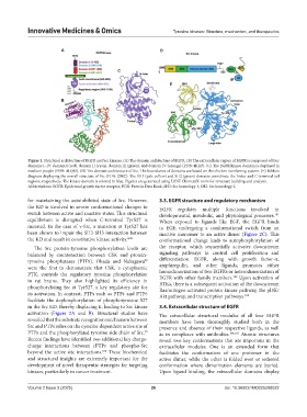
Figure 1. Structural architecture of EGFR and Src kinases. (A) The domain architecture of EGFR. (B) The extracellular region of EGFR is composed of four
domains I–IV: domain I (red), domain II (cyan), domain III (green), and domain IV (orange) (PDB: 4KRP). (C) The EGFR kinase domain is displayed in
medium purple (PDB: 4LQM). (D) The domain architecture of Src. The boundaries of domains are based on the chicken numbering system. (E) Ribbon
diagram displaying the overall structure of Src (PDB: 2SRC). The SH3 (pale yellow) and SH2 (green) domains coordinate the linker and C-terminal tail
regions, respectively. The kinase domain is colored in blue. Figures are generated using UCSF ChimeraX: tools for structure building and analysis.
Abbreviations: EGFR: Epidermal growth factor receptor; PDB: Protein Data Bank; SH3: Src homology 3; SH2: Src homology 2.
for maintaining the autoinhibited state of Src. However, 3.3. EGFR structure and regulatory mechanism
the KD is involved in severe conformational changes to EGFR regulates multiple functions involved in
switch between active and inactive states. This structural developmental, metabolic, and physiological processes.
101
equilibrium is disrupted when C-terminal Tyr527 is When exposed to ligands like EGF, the EGFR binds
mutated. In the case of v-Src, a mutation at Tyr527 has to EGF, undergoing a conformational switch from an
been shown to impair the SH2-SH3 interaction between inactive monomer to an active dimer (Figure 2C). This
the KD and result in constitutive kinase activity. 4,89 conformational change leads to autophosphorylation of
The Src protein-tyrosine phosphorylation levels are the receptor, which sequentially activates downstream
balanced by counteraction between CSK and protein- signaling pathways to control cell proliferation and
tyrosine phosphatases (PTPs). Okada and Nakagawa differentiation. EGFR, along with growth factor-a,
98
were the first to demonstrate that CSK, a cytoplasmic amphiregulin, and other ligands, promotes either
PTK, controls the regulatory tyrosine phosphorylation homodimerization of two EGFRs or heterodimerization of
102
in rat brains. They also highlighted its efficiency in EGFR with other family members. Upon activation of
RTKs, there is a subsequent activation of the downstream
phosphorylating Src at Tyr527, a key regulatory site for Ras/mitogen-activated protein kinase pathway, the pI3K/
its activation. In contrast, PTPs such as PTPε and PTPε Akt pathway, and transcription pathways. 103
facilitate the dephosphorylation of phosphotyrosine 527
in the Src KD, thereby displacing it, leading to Src kinase 3.4. Extracellular structure of EGFR
activation (Figure 2A and B). Structural studies have The extracellular structural modules of all four EGFR
revealed that the substrate recognition mechanism between members have been thoroughly studied both in the
Src and PTPs relies on the cysteine-dependent active site of presence and absence of their respective ligands, as well
99
PTPs and the phosphorylated tyrosine side chain of Src. as in complexes with antibodies. 104,105 Atomic structures
Recent findings have identified two additional key charge- reveal two key conformations that are important in the
charge interactions between rPTPε and phospho-Src extracellular modules. One is an extended form that
beyond the active site interactions. These biochemical facilitates the conformation of one protomer in the
100
and structural insights are extremely important for the active dimer, while the other is folded over or tethered
development of novel therapeutic strategies for targeting conformation where dimerization elements are buried.
kinases, particularly in cancer treatment. Upon ligand binding, the extracellular domains display
Volume 2 Issue 3 (2025) 26 doi: 10.36922/IMO025200022