
Page 8 - IMO-2-3
P. 8
Innovative Medicines & Omics MSC exosomes for digestive tumors: Bench to bedside
tumors of the digestive system includes surgical treatment, Notably, most of the identified exosomal proteins, such as
radiotherapy, and drug therapy. For most patients with heat shock proteins and MHC molecules, are also found
metastatic malignant tumors, curative treatment is often in other types of extracellular vesicles. However, a series of
no longer a viable option. Palliative chemotherapy has proteins is relatively specific to exosomes, including CD9,
been the main systemic drug treatment, but its clinical CD63, CD81, TSG101, Alix, HSP70, and HSP90. These
application is limited by significant toxicity. In recent proteins are considered markers for identifying exosomes.
years, targeted therapy has further improved treatment The lipid composition of exosomes is mainly divided into
efficacy; however, off-target toxicity remains a pressing four categories: sphingolipids, phospholipids, glycolipids,
issue in clinical practice. As research into the mechanisms and fatty acids. Thousands of RNA molecules, including
2
of mesenchymal stem cells (MSCs) and MSC-derived miRNAs, long non-coding RNAs (lncRNAs), and circular
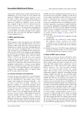
exosomes (MSC-exo) deepens, growing evidence suggests RNAs, have been identified in exosomes. 9-13 The process
that both MSCs and MSC-exo hold great potential in of exosome formation (Figure 1) mainly involves the
treating malignant tumors. 3 following steps:
(i) Endocytosis: The cell membrane invaginates, forming
2. MSCs and their exos the endosome.
2.1. MSCs (ii) Transformation into multivesicular bodies (MVBs):
The endosome further transforms into MVBs.
MSCs represent a class of pluripotent stem cells (iPSCs) During this process, the membrane of the endosome
capable of self-renewal and differentiation into various invaginates, forming multiple small vesicles with lipid
cell types. Under specific induction conditions, MSCs can bilayers.
differentiate into various tissue cells, including adipocytes,
muscle cells, tendon cells, ligament cells, nerves cells, liver (iii) Release of exosomes: The MVBs merge with the cell
cells, cardiomyocytes, and endothelial cells, among others. membrane, discharging their internal small vesicles
14
MSCs typically exhibit spindle-shaped or stellate adherent into the extracellular environment as exosomes.
growth, with high expression of CD73, CD90, and CD105, 3. Roles of MSC-exo in cancer
and low expression of CD34, CD45, CD14 or CD11b,
CD79a or CD19, and HLA-DR. Even after repeated Tumor tissues are made up of tumor cells and the tumor
passage culture and cryopreservation, MSCs retain their microenvironment (TME). Tumor cells, having lost their
multilineage differentiation potential, making them an normal regulatory mechanisms, can grow uncontrollably,
ideal choice for cell therapy. Clinically, MSCs have achieved invade nearby tissues, and spread to distant parts of the
4
significant breakthroughs in treating various diseases, body. The TME includes various components, such as
including hematological diseases, cardiovascular diseases, endothelial cells, T cells, natural killer T-cells, myeloid-
liver cirrhosis, neurological disorders, and autoimmune derived suppressor cells, cancer-associated fibroblasts
5
diseases. More importantly, MSCs can regulate tumor (CAFs), and tumor-associated stromal cells, among
15
growth through various mechanisms. others. MSC-exo plays a crucial role in tumor growth by
transporting regulatory molecules. Interestingly, growing
2.2. Exosome formation and components evidence suggests that MSC-exo can have dual effects
Exosomes are nanoscale vesicles with a closed membrane in cancer, acting as a double-edged sword. Some studies
structure, typically ranging from 40 to 100 nm in diameter, indicate that MSC-exo can promote tumor growth. For
16
and are encapsulated by a lipid bilayer. These vesicles example, Wang et al. demonstrated that exosomes derived
contain many biologically functional molecules – such from bone marrow mesenchymal stem cells (BMMSC-exo)
as proteins, nucleic acids, and lipids – and serve as an from both multiple myeloma patients and healthy donors
important medium for transmitting biological signals can enhance the growth of multiple myeloma (MM) cells
between cells. In 1983, exosomes were discovered for the by activating several signaling pathways related to cell
6
1 time, whereas in 2007, exosomes were found to contain proliferation, such as p38, p53, and Akt. Further research
7
st
microRNAs (miRNA) and mRNAs, confirming that by Deng et al. confirmed that LINC00461 in BMMSC-
17
genetic material can be exchanged between cells through exo from multiple myeloma patients increases BCL-2
exosomes, thereby altering cellular biological behavior. expression by targeting miR-15a/16, thereby preventing
8
Exosomal proteins generally encompass a variety of apoptosis in multiple myeloma cells. Likewise, studies
types, such as integral membrane proteins, peripheral have also reported that MSC-exo inhibits tumor growth.
membrane proteins, outer membrane proteins anchored For instance, BMMSC-exo can release miR-222-3p, which
by lipids, inner membrane proteins anchored by lipids, directly targets the IRF2 gene, thereby negatively regulating
surface proteins, and enzymes associated with exosomes. the IRF2/INPP4B signaling pathway in THP-1 cells and
Volume 2 Issue 3 (2025) 2 doi: 10.36922/IMO025210025