
Page 9 - IMO-2-3
P. 9
Innovative Medicines & Omics MSC exosomes for digestive tumors: Bench to bedside
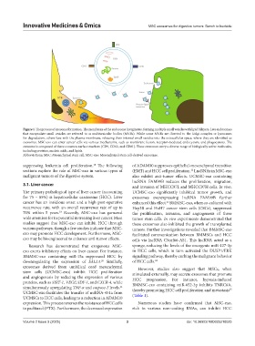
Figure 1. The process of exosome formation. The membrane of the endosome invaginates, forming multiple small vesicles with lipid bilayers. Late endosomes
that encapsulate small vesicles are referred to as multivesicular bodies (MVBs). While some MVBs are directed to the Golgi complex or lysosomes
for degradation, others fuse with the plasma membrane, releasing their internal small vesicles into the extracellular space, where they are identified as
exosomes. MSC-exo can enter cancer cells via various mechanisms, such as membrane fusion, receptor-mediated endocytosis, and phagocytosis. The
exosome is composed of three common surface markers (CD9, CD63, and CD81). These exosomes carry a diverse range of biologically active molecules,
including proteins, nucleic acids, and lipids.
Abbreviations: MSC: Mesenchymal stem cell; MSC-exo: Mesenchymal stem cell-derived exosomes.
18
suppressing leukemia cell proliferation. The following of ADAM10 suppresses epithelial-mesenchymal transition
sections explore the role of MSC-exo in various types of (EMT) and HCC cell proliferation. LncRNAs in MSC-exo
22
malignant tumors of the digestive system. also exhibit anti-tumor effects. UCMSC-exo containing
lncRNA FAM99B reduces the proliferation, migration,
3.1. Liver cancer and invasion of MHCC97L and MHCC97H cells. In vivo,
The primary pathological type of liver cancer (accounting UCMSC-exo significantly inhibited tumor growth, and
for 75 – 85%) is hepatocellular carcinoma (HCC). Liver exosomes overexpressing lncRNA FAM99B further
cancer has an insidious onset and a high post-operative enhanced this effect. BMMSC-exo, when co-cultured with
23
recurrence rate, with an overall recurrence rate of up to Hep3B and HuH7 cancer stem cells (CSCs), suppressed
70% within 5 years. Recently, MSC-exo has garnered the proliferation, invasion, and angiogenesis of these
19
wide attention for its potential in treating liver cancer. Most tumor stem cells. In vivo experiments demonstrated that
studies suggest that MSC-exo can inhibit HCC through these exosomes also inhibited the growth of transplanted
various pathways, though a few studies indicate that MSC- tumors. Further investigations revealed that BMMSC-exo
exo may promote HCC development. Furthermore, MSC- facilitated communication between BMMSCs and HCC
exo may be bioengineered to enhance anti-tumor effects. cells via lncRNA C5orf66-AS1. This lncRNA acted as a
Research has demonstrated that exogenous MSC- sponge, reducing the levels of the oncogenic miR-127-3p
exo exerts inhibitory effects on liver cancer. For instance, in HCC cells, which in turn activated the DUSP1/ERK
BMMSC-exo containing miR-15a suppressed HCC by signaling pathway, thereby curbing the malignant behavior
downregulating the expression of SALL4. Similarly, of HCC cells. 24
20
exosomes derived from umbilical cord mesenchymal However, studies also suggest that MSCs, when
stem cells (UCMSC-exo) inhibit HCC proliferation stimulated externally, may secrete exosomes that promote
and angiogenesis by reducing the expression of various HCC progression. For instance, hypoxia-induced
proteins, such as SIRT-1, VEGF, SDF-1, and CXCR-4, while BMMSC-exo containing miR-652-3p inhibits TNRC6A,
simultaneously upregulating TNF-α and caspase-3 levels. 25
21
UCMSC-exo facilitates the transfer of miRNA-451a from thereby promoting HCC cell proliferation and metastasis
UCMSCs to HCC cells, leading to a reduction in ADAM10 (Table 1).
expression. This process reverses the resistance of HCC cells Numerous studies have confirmed that MSC-exo,
to paclitaxel (PTX). Furthermore, the decreased expression rich in various non-coding RNAs, can inhibit HCC
Volume 2 Issue 3 (2025) 3 doi: 10.36922/IMO025210025