Page 82 - ITPS-7-1
P. 82

INNOSC Theranostics and
            Pharmacological Sciences                                                PfHSP and polyamines interactions


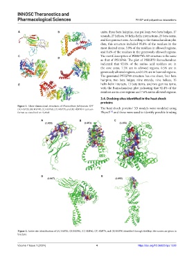
            A                        B                         units, three beta hairpins, one psi loop, two beta bulges, 17
                                                               strands, 27 helices, 36 helix-helix interactions, 25 beta turns,
                                                               and five gamma turns. According to the Ramachandran plot
                                                               data, this structure included 95.8% of the residues in the
                                                               most desired areas, 3.9% of the residues in allowed regions,
                                                               and 0.4% of the residues in the generously allowed regions.
            C                                                  The motif description of PfHSP70’s 3D structure is the same
                                     D
                                                               as that of PfHSP60. The plot of PfHSP70 Ramachandran
                                                               indicated that 92.0% of the amino acid residues are in
                                                               the core areas, 7.3% are in allowed regions, 0.5% are in
                                                               generously allowed regions, and 0.2% are in banned regions.
                                                               The generated PfHSP90 structure has one sheet, four beta
                                                               hairpins, two beta bulges, nine strands, nine helices, 15
            E                                                  helix-helix interacts, 13 beta turns, and two gamma turns,
                                                               with the Ramachandran plot indicating that 92.4% of the
                                                               residues are in core regions and 7.6% are in allowed regions.

                                                               3.4. Docking sites identified in the heat shock
                                                               proteins
            Figure 1. Three-dimensional structures of Plasmodium falciparum 3D7
            (A) HSP20, (B) HSP40, (C) HSP60, (D) HSP70, and (E) HSP90 in cartoon   The heat shock proteins’ 3D models were modeled using
                                                                    [15]
            format as visualized on PyMol.                     Phyre2  and these were used to identify possible binding
                          A                       B                    C


                          D                                    E


            Figure 2. Active site identification of (A) HSP20, (B) HSP40, (C) HSP60, (D) HSP70, and (E) HSP90 identified through SiteMap. Site scores are given in
            brackets.


            Volume 7 Issue 1 (2024)                         4                         https://doi.org/10.36922/itps.1228
   77   78   79   80   81   82   83   84   85   86   87