
Page 8 - JCTR-10-3
P. 8
182 Xie et al. | Journal of Clinical and Translational Research 2024; 10(3): 180-190
for 30 s, with a final extension at 72°C for 5 min. The amplified Quantitative values are expressed as the mean ± standard deviation
products from gallstones, bile, gallbladder mucosa, and feces (M ± SD). Two-sample independent t-test and the Wilcoxon rank-
samples were verified by gel electrophoresis with a 1.5% sum test were used between the two groups. For multi-group
agarose gel, a mixture of 3 μL PCR product and 3 μL 3× loading comparison, one-way analysis of variance and the Kruskal–Wallis
buffer, and 3 μL 100 bp ladder marker (Yingwei Jieji Trading, rank-sum test were used. Statistical significance was set at P < 0.05.
China) at 100V voltage over 35 – 40 min.
Agencourt AMPure XP (Beijing Huaruikang Technology, 3. Results
China) was used to purify the 16S V3-V4 amplicons to be free 3.1. Study population characteristics
of primers and primer-dimer species. The second PCR reaction
was performed in a 25 μL mixture containing 5 μL 5× GC This study investigated the relationship between gallstone
buffer, 0.75 μL KAPA dNTP mix, 0.5 μL KAPA HiFi HotStart formation and bacteria in the bile, gallbladder mucosa, and
DNA polymerase, 1.5 μL barcode F (10 pM), 1.5 μL barcode R feces of 21 gallstone patients (eight males and 11 females; age
(10 pM), 5 μL purified product, and 10.75 μL retinoblastoma. range: 32 – 73 years old). From the gallstone group, we obtained
The purified product was amplified by PCR using primers, 13 gallstone specimens (S1 – S13), nine bile specimens (Z1 –
where the barcode is an eight-base sequence unique to each Z9), 13 gallbladder mucosa specimens (N1 – N13), and 17 feces
sample. Denaturation, annealing, elongation, and cycling were specimens (F1 – F17). Meantime, we collected 20 feces (HF1
the same as the first PCR amplification. The amplicons were – HF17) samples from the control group. We rejected three
subsequently purified by AMPure XP beads to clean up the final samples due to amplification failure; one from the gallstone
library before quantification. Finally, purified amplicons were specimens, one from the gallstone patients’ feces specimens, and
pooled in equimolar and paired-end sequences (2 × 250) on an one from the healthy subjects’ feces specimens. The average age
Illumina MiSeq platform according to the standard protocols. and BMI of the patients in the gallstone group were higher than
that of the control group (P = 0.004). There were no statistically
2.4. Bioinformatics analysis of sequencing data significant differences in gender and cholesterol levels between
the gallstone and control groups (Table 1).
Fast length adjustment of short reads was used to merge
paired-end reads from next-generation sequencing [15]. Low- 3.2. Bacterial diversity of sample species under different
quality reads were filtered by fastq_quality_filter (−p 90 −q sequencing quantities and OTUs dilution curve
25 −Q 33) in FASTX Toolkit 0.0.14, and chimera reads were In this study, we identified a total of 23427 OTUs (340 ±
removed by USEARCH 64-bit version 8.0.1517. The number of 93) based on the conventional criterion of 97% sequence
reads for each sample was normalized based on the smallest size similarity, with 4095 OTUs in gallstones, 3065 OTUs in bile,
of samples by random subtraction. The final optimized sequence 4687 in gallbladder mucosa, 5203 OTUs in patients’ feces, and
was obtained to ensure the reliability of the effective sequence 6377 OTUs in normal feces. There was no significant difference
used as operational taxonomic units (OTUs). OTUs were aligned in the intestinal microbiota diversity based on the feces of the
by the Uclust algorithm with a 97% identity and taxonomically gallstone and control groups. There was also no statistical
classified using the Silva16S rRNA database (https://www. difference in the bacterial diversity between gallstones, bile, and
arbsilva.de/documentation/release-128/). From the levels of gallbladder mucosa in the gallstone group (P > 0.05). The gut
phylum and genus, the dominant bacteria obtained by sequencing microbiota was reportedly diverse in gallstones (P = 0.004), bile
in each group were statistically analyzed. The α-diversity reflects a (P = 0.045), and gallbladder mucosa (P = 0.008). In addition,
comprehensive indicator of microbial evenness and abundance in the gut microbiota was more diverse in the gallstone group than
a single sample and mainly includes the abundance index Chao1, the control group (Table 2).
Shannon’s index, and Simpson’s index. In contrast, β-diversity When the number of sequences increased, the diversity
is a comparative analysis of microbial community composition index did not increase significantly, indicating that the number
among different groups. Both α- and β-diversities were generated of sequences was sufficient to reflect the overall community
in the Quantitative Insights Into Microbial Ecology (QIIME) structure (Figure 1). In addition, the increase in the number of
software and calculated based on weighted and unweighted sequences did not generate new OTUs.
Unifrac distance matrices [16]. Venn diagram selects OTUs
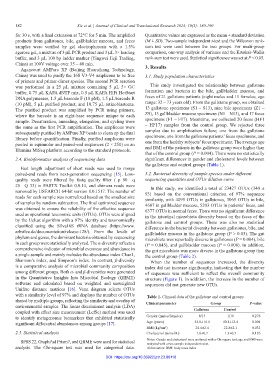
with a similarity level of 97% and displays the number of OTUs Table 1. Clinical data of the gallstone and control groups
shared by multiple groups, reflecting the similarity and overlap of Clinical parameter Group P‑value
environmental samples. The linear discriminant analysis (LDA)
coupled with effect size measurement (LefSe) method was used Gallstone Control
to identify metagenomic biomarkers that exhibited statistically Gender (males/females) 8/13 11/9 0.278
significant differential abundances among groups [17]. Age (years) 52.8±14.4 40.1±12.4 0.004
BMI (kg/cm ) 24.4±2.4 22.8±2.1 0.032
2
2.5. Statistical analysis Cholesterol (mmol/L) 1.6±0.7 1.3±0.5 0.126
SPSS 22, GraphPad Prism7, and QIIME were used for statistical Notes: Gender and cholesterol were analyzed with a Chi-square test; age and BMI were
analyzed with a two-sample independent t-test.
analysis. The Chi-square test was used for categorical data. Abbreviation: BMI: body mass index
DOI: https://doi.org/10.36922/jctr.23.00118