
Page 30 - JCTR-10-4
P. 30
252 Sari et al. | Journal of Clinical and Translational Research 2024; 10(4): 246-255
A B
C D E
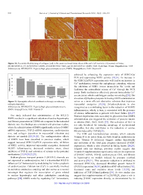
Figure 10. Picrosirius Red staining of collagen (red) in the cross-sectional tissue slices of the rat’s left ventricle: (A) normal rat tissue;
(B) HF/HG/STZ; (C) HF/HG/STZ+EMPA; (D) HF/HG/STZ+VitD; and (E) HF/HG/STZ+EMPA+VitD. Scale bars: 50 µm. Magnification: ×400
Abbreviations: HF/HG/STZ: High-fat/high-glucose/streptozotocin; EMPA: Empagliflozin; VitD: Vitamin D
achieved by enhancing the expression ratio of SERCA2a/
PLN and suppressing NHE1 activity [19,24]. An increase in
the SERCA2a/PLN expression ratio will lead to an increase in
Ca mobilization toward the endoplasmic reticulum, whereas
2+
the inhibition of NHE1 lowers intracellular Na levels and
+
facilitates the extracellular release of Ca through the NCX
2+
pump. Both mechanisms effectively prevent intracellular Ca
2+
accumulation which could trigger cardiac remodeling [25]. The
elevation of β-hydroxybutyrate following EMPA administration
Figure 11. Synergistic effect of combination therapy on reducing serves as a more efficient alternative substrate that improves
collagen deposition myocardial energetics [15,24]. β-hydroxybutyrate is also
Abbreviations: HF/HG/STZ: High-fat/high-glucose/streptozotocin; recognized as a contributing factor to the reduction of NLRP3
EMPA: Empagliflozin; VitD: Vitamin D inflammasome, which, in turn, is associated with the presence
of chronic inflammation in patients with heart failure [26,27]
Our study indicated that administration of the SGLT-2i Nutrient deprivation state secondary to glycosuria from EMPA
EMPA resulted in a significant reduction of cardiac hypertrophy administration also triggered the activation of proteins known
and fibrosis parameters in T2DM rats compared to the untreated as sirtuins (Sirt1, Sirt3, Sirt6) [23]. The activation of Sirt1 is
diabetic rats. Our findings are consistent with previous studies, not only beneficial for inducing autophagy of dysfunctional
where the administration of EMPA significantly reduces β-MHC organelles, but also contributes to the reduction of cardiac
mRNA expression, TGF-β mRNA expression, cardiomyocyte fibrosis induced by TGF-β [23].
size, and collagen deposition in myocardial infarction and The VDR and 1-α-hydroxylase enzyme, which converts
diabetic rat models [15,19-21]. The cardioprotective effects Vitamin D to its active form, are both found in cardiovascular
of EMPA have been described in previous studies, which tissue. Vitamin D deficiency triggers cardiac hypertrophy
suggest increased SERCA2a/PLN expression ratio, inhibition and activation of the fetal gene program (increased β-MHC
of NHE1 activity, improved myocardial energetics, decreased expression), which is also observed in failing hearts [28,29].
NLRP3 inflammasome, decreased oxidative stress, direct Aligned with our findings, previous studies suggest that
inhibition of TGF-β, and activation of sirtuins as the potential Vitamin D supplementation significantly reduces the expression
mechanisms [15,19,20,22,23]. of β-MHC mRNA, TGF-β, cardiomyocyte CSA, and fibrosis
Sodium-glucose transport protein 2 (SGLT-2) channels are in hypertrophic rat models induced by pressure overload
not expressed in cardiomyocytes, but it is known that SGLT-2i and uremia [30,31]. This parameter reduction is attributed to
influences Ca homeostasis by modulating Na in the cytoplasm increased SERCA2a, decreased fibroblast growth factor-23
+
2+
of cardiomyocytes. Calcium ion (Ca ) is essentially involved (FGF23) expression [30,31], inhibition of NF-κB activation [32],
2+
in excitation-contraction coupling and also serves as a second decreased renin and oxidative stress levels [13,33], and
messenger that regulates the transcription of genes related inhibition of TGF-β/Smad pathway [34]. In vitro studies also
to cardiac hypertrophy and other maladaptive remodeling suggest that supplementation of 1α,25(OH) D plays a role in
3
2
pathways [24]. EMPA works by regulating Ca homeostasis, reducing β-MHC expression directly, as demonstrated by its
2+
DOI: http://doi.org/10.36922/jctr.24.00010