
Page 49 - JCTR-10-5
P. 49
Arnold and Arm | Journal of Clinical and Translational Research 2024;10(5):307-316 311
In this case, a 65-year-old male presented with a poorly 28% of global chronic wounds that require treatment, accounting
granulated 1 cm left plantar Charcot DFU. The wound had for nearly $2 billion in annual expenditures, representing an
2
been open for more than 1 year despite standard care, including enormous and growing global problem [28]. Up to 60% of
serial debridement, negative pressure wound therapy (NPWT), VLUs are considered chronic because they persist for more
and total contact casting. The lesion extended 0.4 cm deep to than 6 weeks, usually as a result of blood circulation problems.
devitalized bone, and the patient was being treated concurrently Obesity, smoking, deep vein thrombosis (DVT), varicose veins,
for chronic osteomyelitis on presentation and initiation of previous leg injury or surgery, age, and diabetes are all risk
PMVT treatment. Between 2 and 4 weeks, offloading non- factors that can contribute to the development of a VLU [28].
compliance led to re-emergence of infection, requiring As PMVT is intended to improve blood flow through the repair
significant debridement and initiation of antibiotics. Following and reconstruction of microvascular tissue by serving as a
this, the wound area was now 24 cm . Despite this setback, after scaffold for cellular invasion and capillary growth, the benefits
2
three additional PMVT applications, the wound had become of improved microcirculatory blood flow may be particularly
99% epithelialized, and PMVT treatment was discontinued. impactful on patients with compromised microvasculature, such
The ulcer went on to fully close within 6 additional weeks as patients with increased risk for non-healing VLUs.
with standard care and has remained healed 6 months to date The 64-year-old male with a history of chronic kidney disease
following closure. Images of the ulcer progression and the (Stage 3b), hypertension, chronic DVT, Leiden Factor V, and
wound area/volume graph are displayed in Figure 3A and B. asthma, who presented with a left VLU in August 2021, is an
example of such a patient. The ulcer worsened following DVT in
3.3. Case 3: Increasing blood flow using PMVT to treat a April 2022. No thrombectomy interventions were advisable due
chronic venous leg ulcer
to an unacceptably high risk of complications. Multiple topical
Venous leg ulcers (VLUs), among the most common types of wound management products, including silver alginate, foam,
lower extremity wounds, are open ulcers that frequently occur hydroconductive and other composite dressings, cadexomer
on the inside of the leg above the ankle. VLUs comprise about iodine, topical antibiotics, and an ECM xenograft, along with
compression wraps (the patient’s job requires standing 8 h a
day), all failed to close the VLU.
A
In January 2024, after nearly 2.5 years of not healing, the
patient received his first treatment of PMVT. The PMVT disk was
removed from the vial and applied directly onto the surface of the
ulcer after very minimal selective debridement. On contact with
blood at the wound site, the PMVT graft was quickly absorbed
into the surrounding tissue. Two additional PMVT applications
were made at weeks 1 and 5 after the initial treatment. A non-
adherent dressing (Mepitel; Mölnlycke Health Care, USA) was
used to cover 1 – 2 cm beyond the ulcer’s edges after each PMVT
application. The VLU was covered further by a compression
bandage (initially an alginate-based absorbent compress for
B the first two treatments, then Coban 2 Lite [3M Healthcare,
USA] for the final application). The patient suffered a right hip
fracture requiring surgery and hospitalization 2 weeks after initial
treatment and had limited transportation means for further follow-
up visits until week 6, resulting in a gap between the second and
third PMVT applications. As with the other cases, the VLU was
visually examined and photographed at each visit, and the wound
size was measured using a ruler. In addition, tissue oxygenation
was assessed using a point-of-care near-infrared (NIR) imaging
device (Snapshot NIR; Kent Imaging, Canada).
The ulcer closed after just three applications of PMVT.
Images of the VLU’s progression are displayed in Figure 4A,
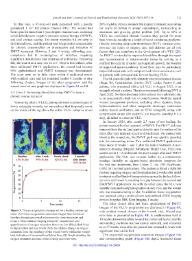
Figure 5. Tissue oxygenation changes within a healing venous leg with evident wound closure at the 6-week visit. Wound size
ulcer. (A) Tissue oxygenation saturation images from historical over time is presented in Figure 4B. A confirmation visit at
baseline through processed microvascular tissue treatment and 10 weeks demonstrated the wound had remained healed and the
closure. Near-infrared imaging allows for visualization and
quantification of oxygen saturation from very low (black/dark blue) surrounding erythema had noticeably subsided, and remained
to high (yellow and red) levels. Note the relative change in oxygen so at 17 weeks, even after the patient had returned to work with
saturation from the periphery of the wound bed to within the wound significant time on his feet.
bed, indicative of increased local blood flow. (B) Graph detailing the The sequential oxygenation saturation images (Figure 5A)
oxygen saturation increase of the closing ulcer over time. and corresponding graph (Figure 5B) depict increased tissue
DOI: http://doi.org/10.36922/jctr.24.00059