
Page 50 - JCTR-10-5
P. 50
312 Arnold and Arm | Journal of Clinical and Translational Research 2024;10(5):307-316
oxygenation within the center region of the wound area from the surgical skin defect to be at greater risk for not healing. In such
historical baseline (13%), after initial PMVT treatment (49%), cases, proactive use of an advanced wound care treatment may
through healing just before closure (62%), and maintained be warranted.
following closure (61%). Although these numbers are relative, The first Mohs patient, a 51-year-old male with coronary
the increased oxygen saturation represents improved blood artery disease, hypertension, nicotine dependence, and post-
flow and is indicative of the transition to the proliferative and COVID pulmonary issues, had undergone Mohs excision of
remodeling phases of healing within the wound area. a basal cell carcinoma on his right scapula. Initial attempts to
By repairing the deficient local microvasculature around the close the defect using standard treatment and negative pressure
VLU, PMVT was able to assist with the delivery of oxygen wound therapy were unsuccessful, and he presented 5 weeks
and nutrients to the ulcer. With just three topical applications, it post-excision with a defect 13 cm in area and 0.4 cm deep. After
2
successfully healed a challenging ulcer that had not closed after just one treatment of topical PMVT, over 50% of the wound
over 2.5 years of conventional wound management. volume had been replaced with new tissue. After 5 weekly
applications, the defect had closed, and PMVT treatment was
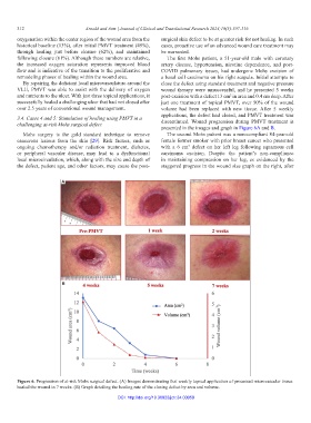
3.4. Cases 4 and 5: Stimulation of healing using PMVT in a discontinued. Wound progression during PMVT treatment is
challenging at-risk Mohs surgical defect
presented in the images and graph in Figure 6A and B.
Mohs surgery is the gold standard technique to remove The second Mohs patient was a non-compliant 84-year-old
cancerous lesions from the skin [29]. Risk factors, such as female former smoker with prior breast cancer who presented
ongoing chemotherapy and/or radiation treatment, diabetes, with a 6 cm defect on her left leg following squamous cell
2
or peripheral vascular disease, may lead to a dysfunctional carcinoma excision. Despite the patient’s non-compliance
local microcirculation, which, along with the size and depth of in maintaining compression on her leg, as evidenced by the
the defect, patient age, and other factors, may cause the post- staggered progress in the wound size graph on the right, after
A
B
Figure 6. Progression of at-risk Mohs surgical defect. (A) Images demonstrating that weekly topical application of processed microvascular tissue
healed the wound in 7 weeks. (B) Graph detailing the healing rate of the closing defect by area and volume.
DOI: http://doi.org/10.36922/jctr.24.00059