
Page 50 - JCTR-11-1
P. 50
Journal of Clinical and
Translational Research Hesperidin enhances repair of γ-irradiated wounds
A B
C
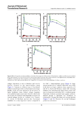
Figure 8. Effect of hesperidin on lipid peroxidation in the skin of mice exposed to different doses of fractionated γ- radiation at different post-irradiation
times. MDA levels in (A) 10 Gy, (B) 20 Gy, and (C) 40 Gy. Notes: Squares indicates: CMC+Sham-irradiation; Uptriangles indicates: Hesperidin+Sham-
irradiation; Circles indicates: CMC+irradiation; Down triangles indicates: Hesperidin+irradiation.
Abbreviations: CMC: Carboxymethylcellulose; HPD: Hesperidin; IR: Irradiation; MDA: Malondialdehyde; SIR: Sham-irradiation.
collagen deposition as well as fibroblast and vascular the CMC + sham-irradiation group (Figure 9). Large,
densities compared to the sham-irradiation group unusual stellate cells or “radiation fibroblasts” were visible
(Figure 9). Exposure to different doses of fractionated in the dermis at higher radiation doses, especially at 20
radiation caused degeneration and hyalinization of collagen and 40 Gy. Epidermal thickness varied with increasing
bundles, which was more apparent at 20 and 40 Gy. At radiation dose, and irregularly shaped blood vessels were
higher irradiation doses, isolated collagen fragments seen in all exposure doses in the CMC + irradiation group.
surrounded by empty spaces were observed, which were Hesperidin treatment protected mice against radiation-
not stained. Pre-treatment with hesperidin increased and induced damage to fibroblasts and vasculature, as
restored collagen bundles in irradiated mice. However, evidenced by increased fibroblasts and vascular densities
the density of fibroblasts declined after fractionated (Figures 9 and 10). However, the normal histological
irradiation in a dose-dependent manner compared to picture was not restored.
Volume 11 Issue 1 (2025) 44 doi: 10.36922/jctr.24.00049