
Page 69 - JCTR-11-2
P. 69
Journal of Clinical and
Translational Research Fetal posterior fossa imaging findings
Transabdominal ultrasound, a key component of fetal
imaging, is complemented by fetal magnetic resonance
imaging (MRI) as a crucial adjunct. Guidelines from
reputable organizations such as American College of
Obstetricians and Gynecologists (ACOG), American
Institute of Ultrasound in Medicine (AIUM), Australasian
Society for Ultrasound in Medicine (ASUM), National
Health Service (NHS), and International Society of
Ultrasound in Obstetrics and Gynecology (ISUOG)
emphasize the importance of posterior fossa assessment
during second-trimester pregnancy imaging. MRI,
3-7
when performed without contrast media, has no known
adverse fetal effects at any stage of pregnancy. Over the
7
past decade, MRI has improved prenatal diagnosis of CNS
8
anomalies, especially in the posterior fossa. Despite some
controversy in terminology, diagnosing posterior fossa Figure 1. The fetal rhombencephalon, observed as an anechoic structure
malformations can significantly aid parental counseling in the posterior brain (yellow arrow), is a normal finding at 8 – 10 weeks
and pregnancy management. of gestation and is not indicative of developmental issues.
Abbreviation: GA: Gestational age.
The primary aim of this review is to evaluate and
compare the roles of ultrasound and MRI in diagnosing
fetal posterior fossa anomalies, with a focus on cerebellar
malformations. It seeks to clarify diagnostic criteria,
address ambiguous terminology, and propose standardized
protocols that could improve parental counseling and
clinical decision-making.
2. Methods
A comprehensive literature search was conducted using
PubMed to identify relevant articles published up to
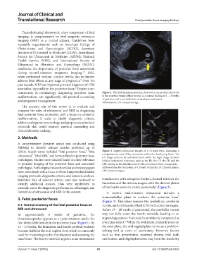
(2024). Search terms included “fetal posterior fossa,” “fetal Figure 2. Sagittal ultrasound images of a 13-week fetus, displaying a
ultrasound,” “fetal MRI,” and terms related to posterior fossa comprehensive view of key structures within the developing brain. The
left image presents an unmarked scan, while the right image includes
pathologies. Studies were selected based on their relevance labelled anatomical structures, such as the BS, the 4.V, the CP, and the
to prenatal imaging of the posterior fossa and associated CM, helping in the identification of critical posterior fossa components.
pathologies. Both original research articles and review papers Abbreviations: BS: Brainstem; 4.V: Fourth ventricle; CP: Choroid plexus;
were considered, with a focus on those that provided detailed CM: Cisterna magna.
imaging protocols, diagnostic criteria, and outcome analyses.
Reference lists of selected articles were also reviewed to translucency with echogenic borders, located between the
identify additional sources. Data were synthesized to brainstem and the cisterna magna, with the choroid plexus
9
critically assess the diagnostic performance, advantages, and of the fourth ventricle visible posteriorly (Figure 2).
limitations of ultrasound and MRI in this context. A routine mid-trimester ultrasound includes a
7
3. Fetal posterior fossa transcerebellar plane to evaluate the posterior fossa
(Figure 3). This plane assesses the cerebellum, cerebellar
3.1. Normal anatomy of the fetal posterior fossa on vermis, and cerebrospinal fluid (CSF) in the cisterna magna.
MRI and ultrasound Before 19 – 20 weeks of gestational, the cerebellar vermis
At approximately 8 weeks of gestation, the may not fully cover the fourth ventricle, leading to an
rhombencephalon appears as a cystic structure and is the atypical appearance that could be mistakenly interpreted as
10
first identifiable feature in the posterior fossa (Figure 1). By a vermian defect. While the evaluation primarily relies on
11 – 13 weeks, the brainstem and fourth cerebral ventricle the axial plane, the mid-sagittal plane serves as a problem-
become visible in the mid-sagittal view, which is commonly solving tool in cases of uncertainty. However, factors
used for measuring nuchal translucency and assessing the such as fetal presentation, maternal obesity, fetal skull
nasal bone. The fourth ventricle appears as an intracranial ossification, and oligohydramnios may limit the feasibility
Volume 11 Issue 2 (2025) 63 doi: 10.36922/jctr.6240