
Page 74 - JCTR-11-2
P. 74
Journal of Clinical and
Translational Research Fetal posterior fossa imaging findings
A B A B
C
C D
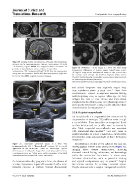
Figure 10. Imaging of mega cisterna magna. (A) Axial ultrasound image
demonstrates the measurement of an enlarged cisterna magna. (B) Axial
TRUFI and (C) Sagittal HASTE MRI images illustrate a n increased CSF Figure 12. Multiplanar TRUFI images of a fetus. (A) Axial image
posterior to the cerebellum, with no evidence of mass effect. demonstrates obliteration of cisterna magna (curved black arrow).
Abbreviations: CSF: Cerebrospinal fluid; TRUFI: True fast imaging with (B) Sagittal image shows herniation of posterior fossa contents into
steady-state free precession; HASTE: Half-Fourier acquisition single-shot the cervical canal through the foramen magnum (black arrow).
turbo spin-echo; MRI: Magnetic resonance imaging.
(C and D) Axial and sagittal images of the torso show a meningomyelocele
sac containing neural tissue (black star).
A B Abbreviations: TRUFI: True fast imaging with steady-state free precession.
and callosal dysgenesis may negatively impact long-
term ambulatory status in some cases. Given these
43
considerations, optimal management requires lifelong
multidisciplinary care, as regular follow-ups can help
mitigate the risks of renal disease and shunt-related
C D complications. In addition, some cases of cardiopulmonary
arrest may be preventable, as they could be linked to Chiari
II malformation decompensation. 44
3.3.8. Occipital encephalocele
An encephalocele is a congenital defect characterized by
E the protrusion of meninges, CSF, and brain tissue through
a cranial defect. These anomalies are categorized based
on their location and can be either open or covered by
skin. Most congenital encephaloceles are associated
with intracranial abnormalities. They may occur as
45
isolated anomalies or as part of syndromes, chromosomal
abnormalities, single-gene disorders, or due to teratogenic
influences. 46
Figure 11. Multiplanar ultrasound images of a fetus with Encephaloceles involve a bony defect in the skull and
meningomyelocele. (A) A “lemon-shaped” cranium. (B) A curved varying degrees of brain tissue displacement (Figure 13).
appearance of the cerebellum, known as “banana-sign” (yellow Imaging shows diverse characteristics, including
arrowhead). (C-E) Ultrasound images show a skin defect (yellow arrow),
a meningomyelocele sac (yellow star), and the separation of vertebral abnormal cortical folding, calcification, and hemorrhage
ossification centers (curved black arrow), respectively. within the externalized brain tissue. In severe cases,
brainstem abnormalities, such as posterior bending
the lesion remains a key prognostic factor, the absence of and atypical configurations, may be present. Surgical
47
vermian displacement is generally associated with a more intervention remains the primary treatment option.
favorable outcome. In contrast, vermian displacement Despite advancements in surgical management, morbidity
Volume 11 Issue 2 (2025) 68 doi: 10.36922/jctr.6240