
Page 76 - JCTR-11-2
P. 76
Journal of Clinical and
Translational Research Fetal posterior fossa imaging findings
A B A B
C
C D
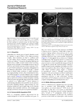
Figure 14. Imaging of rhombencephalosynapsis. (A) Coronal ultrasound Figure 15. Imaging of congenital aqueductal stenosis. Severe
image of a 30-week fetus demonstrates fusion of the cerebellar ventriculomegaly is shown in (A) axial ultrasound and (B) axial HASTE
hemispheres (black arrow). Similar to MRI, the transverse folding of image (white and black star). (C and D) The obstruction at the level of
the fused cerebellum is a distinctive feature. (B) Axial TRUFI image the cerebral aqueduct (black arrow) is visualized in sagittal HASTE and
demonstrates fused cerebellar hemispheres and continuous cerebellar coronal TRUFI images.
folia (curved black arrow). (C) Sagittal TRUFI image shows midline Abbreviations: TRUFI: True fast imaging with steady-state free precession;
sections revealing the absence of the vermis. HASTE: Half-Fourier acquisition single-shot turbo spin-echo.
Abbreviations: MRI: Magnetic resonance imaging; TRUFI: True fast
imaging with steady-state free precession.
date, over twenty types have been identified, each linked
to distinct genetic mutations. While MRI remains the
64
3.3.11. Ciliopathies
primary imaging method, ultrasound can also be used.
Ciliopathies are a diverse group of genetic disorders caused Sagittal imaging plays a crucial role in distinguishing
by mutations in genes linked to the cilium-centrosome cerebellar hypoplasia from pontocerebellar hypoplasia
53
complex. These mutations can lead to conditions such (Figure 17). A mid-sagittal view can show a non-lobulated
as cystic kidney disease, blindness, neurological deficits, cerebellar vermis. The “dragonfly pattern” in a coronal view
59
obesity, and diabetes. Among the ciliopathies affecting the shows flattened cerebellar hemispheres with a relatively
posterior fossa, notable examples include Joubert syndrome, preserved vermis, which resembles the wings and head of
Meckel-Gruber syndrome, orofaciodigital syndrome type VI a dragonfly. In contrast, the “butterfly pattern” indicates a
(OFD-6), and COACH syndrome. Joubert syndrome, OFD- small but proportionate cerebellum. Axial MRI scans of the
6, and COACH syndrome are particularly distinguished by lower mesencephalon often show a “Figure 8” appearance,
the distinctive characteristic “molar tooth sign,” a feature while dilated interfolial spaces suggest a reduction
absent in Meckel-Gruber syndrome. In Joubert syndrome, in parenchymal volume. Although pontocerebellar
65
key imaging findings include a deep interpeduncular fossa, hypoplasia is the primary feature, its clinical manifestation
VH, thickened superior cerebellar peduncle, and an enlarged varies depending on the affected gene, ranging from
fourth ventricle (Figure 16). When additional abnormalities, neurological disorders to joint contractures. Accurate
60
such as cephaloceles and tectocerebellar dysraphia, including classification of PCH subtypes is essential for determining
DWM, are present alongside the classic features, the condition prognosis and guiding neurodevelopmental care. 66
is referred to as “Joubert plus.” 61,62 Recent advances in
understanding ciliopathies have paved the way for precision 3.3.13. Walker-Warburg syndrome (WWS)
treatments, including antisense oligonucleotides for exon WWS is a lethal form of congenital muscular dystrophy
skipping, translational read-through drugs, small molecules that is associated with hydrocephalus, cobblestone
targeting cilium-dependent pathways, and emerging gene lissencephaly, and retinal dysplasia. WWS stands out
67
therapies, all of which show promise in preclinical models. 63 noticeably on MRI due to its unique or characteristic
appearance. The brainstem typically exhibits hypoplasia
3.3.12. Pontocerebellar hypoplasia (PCH) and a “kinked” or “Z” configuration, while the tectum
PCH is a congenital condition characterized by the demonstrates enlargement. Furthermore, the cerebellum
underdevelopment of both the cerebellum and pons. To presents as both small and dysmorphic, with abnormal
Volume 11 Issue 2 (2025) 70 doi: 10.36922/jctr.6240