
Page 79 - JCTR-11-2
P. 79
Journal of Clinical and
Translational Research Fetal posterior fossa imaging findings
A B C
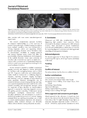
Figure 21. Multimodal imaging of a posterior fossa extra-axial mass-like lesion. (A) Axial HASTE and (B) TRUFI images demonstrate a posterior fossa
extra-axial mass-like lesion with signal characteristics resembling G-ray matter (black star). (C) An ultrasound image confirms the presence of the same
extra-axial lesion across different imaging modalities (yellow arrow).
Abbreviations: HASTE: Half-Fourier acquisition single-shot turbo spin-echo; TRUFI: True fast imaging with steady-state free precession.
often correlate with more severe neurodevelopmental 5. Conclusion
impairments.
Ultrasound and MRI play complementary roles in
This review’s comprehensive approach facilitates diagnosing fetal posterior fossa anomalies. While
an integrated understanding of a wide spectrum of ultrasound is the primary screening tool, MRI enhances
posterior fossa pathologies. Detailed imaging descriptions accuracy when ultrasound is limited. Standardized
across multiple modalities have been instrumental in protocols and multidisciplinary collaboration are essential
refining diagnostic criteria and supporting effective for improving diagnostic consistency and refining prenatal
prenatal counseling. However, several limitations must counseling and management strategies.
be acknowledged. Variability in imaging protocols
and operator expertise across centers may affect the Acknowledgments
generalizability of findings. Furthermore, the heterogeneity We sincerely appreciate Dr. Hasan Yiğit for his assistance
of the available literature—where some conditions are in providing MRI reports, which significantly contributed
described only in isolated case reports or small series— to the study.
coupled with a persistent lack of standardized diagnostic
criteria in certain areas, poses significant challenges to the Funding
overall synthesis of evidence.
None.
Further research is needed to establish standardized
imaging protocols and diagnostic criteria, particularly Conflict of interest
for conditions with overlapping features, such as DWM The authors declare that they have no conflicts of interest.
and VH. Prospective multicenter and multidisciplinary
studies would be instrumental in validating diagnostic Author contributions
measures, exploring advanced imaging techniques,
and fostering a common language among professionals Conceptualization: Deniz Delibaş,
across specialties. Moreover, investigations into the Data curation: Arzu Gülşah Yalçın, Zafer Yumak
correlation between prenatal imaging findings and long- Investigation: Deniz Delibaş, Arzu Gülşah Yalçın, Zafer
term neurodevelopmental outcomes could further refine Yumak
prenatal counseling and management strategies. Given Methodology: Deniz Delibaş
the complexity of these disorders, an interdisciplinary Supervision: Elif Ergün
approach is essential for improving diagnostic accuracy Writing–original draft: Deniz Delibaş
and guiding parental counseling. Collaboration among Writing–review & editing: Elif Ergün
neurologists, radiologists, maternal-fetal medicine Ethics approval and consent to participate
specialists, geneticists, and neonatologists is crucial for
establishing standardized protocols for prenatal imaging, Ethical approval for the collection of clinical data from
postnatal follow-up, and long-term patient care. A well- patients was obtained from the Scientific Research Ethics
structured, multidisciplinary framework will streamline Committee of the TC Sağlık Bakanlığı Ankara Eğitim ve
diagnosis, refine treatment strategies, and ultimately Araştırma Hastanesi (Approval number: E-93471371 –
enhance clinical decision-making and patient outcomes. 514.10 – 255038351, Approval date: September 25, 2024).
Volume 11 Issue 2 (2025) 73 doi: 10.36922/jctr.6240