
Page 78 - JCTR-11-2
P. 78
Journal of Clinical and
Translational Research Fetal posterior fossa imaging findings
A B
C D E
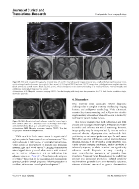
Figure 19. MRI and ultrasound images of a 22-week fetus. (A and B) Axial ultrasound images demonstrate a small cerebellum (yellow dotted lines),
ventriculomegaly, and cobblestone lissencephaly (black arrow). (C-E) Sagittal TRUFI and axial HASTE images show kinked pontomesencephalic junction
(black arrow) and a small vermis (black outlined arrow), which correspond to the ultrasound findings of a small cerebellum, ventriculomegaly, and
cobblestone lissencephaly (black curved arrow).
Abbreviations: MRI: Magnetic resonance imaging; TRUFI: True fast imaging with steady-state free precession; HASTE: Half-Fourier acquisition single-
shot turbo spin-echo.
A B 4. Discussion
Fetal posterior fossa anomalies present diagnostic
challenges due to complex anatomy, overlapping imaging
features, and ambiguous terminology. While ultrasound
remains the primary screening tool, MRI provides valuable
supplementary information when ultrasound is limited by
technical or patient-related factors.
Figure 20. MRI characterization of subacute cerebellar hemorrhage is This review indicates that both ultrasound and MRI
more convient. (A) Axial T1 and (B) coronal TRUFI images show a right possess distinct diagnostic strengths. Ultrasound is widely
cerebellar hematoma (black open arrow) involving the vermis.
Abbreviations: MRI: Magnetic resonance imaging; TRUFI: True fast accessible and effective for initial screening; however,
imaging with steady-state free precession. image quality may be compromised by factors, such as
maternal obesity, oligohydramnios, unfavorable fetal
While most fetal brain tumors occur in supratentorial positioning, or advanced gestational age. In such cases,
regions, posterior fossa mass lesions are less common. One MRI offers superior soft-tissue contrast and multiplanar
71
such pathology is heterotopia, or neuroglial hamartomas, imaging, making it an invaluable complementary tool.
which consist of disorganized yet mature cells, including Under optimal imaging conditions, neither modality is
neurons, glia, and blood vessels. Imaging demonstrates inherently superior, yet their combined use significantly
72
mixed signals from gray and white matter, with minimal enhances overall diagnostic accuracy, ensuring a
or no contrast enhancement and no infiltration into more comprehensive assessment of posterior fossa
adjacent structures (Figure 21). These lesions do not grow structures. Prognosis varies depending on the underlying
over time. Resection is the recommended management etiology and associated anomalies. Isolated cerebellar
73
approach, and the overall prognosis following resection is malformations generally have more favorable outcomes,
excellent, with normal neurological development. 74 whereas additional structural or genetic abnormalities
Volume 11 Issue 2 (2025) 72 doi: 10.36922/jctr.6240