
Page 73 - JCTR-11-2
P. 73
Journal of Clinical and
Translational Research Fetal posterior fossa imaging findings
A A B
C
B C
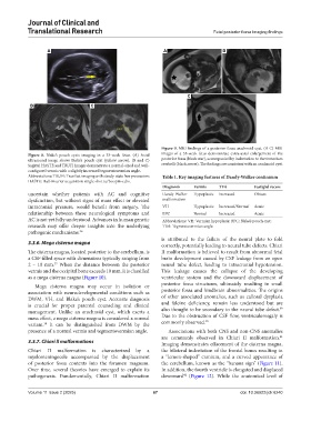
Figure 9. MRI findings of a posterior fossa arachnoid cyst. (A-C) MRI
Figure 8. Blake’s pouch cysts imaging in a 33-week fetus. (A) Axial images of a 35-week fetus demonstrate extra-axial enlargement of the
ultrasound image shows Blake’s pouch cyst (yellow arrow). (B and C) posterior fossa (black star), accompanied by indentation to the tentorium
Sagittal HASTE and TRUFI images demonstrate a normal-sized and well- cerebelli (black arrow). The findings are consistent with an arachnoid cyst.
configured vermis with a slightly increased tegmentovermian angle.
Abbreviations: TRUFI: True fast imaging with steady-state free precession; Table 1. Key imaging features of Dandy‑Walker continuum
HASTE: Half-Fourier acquisition single-shot turbo spin-echo.
Diagnosis Vermis TVA Fastigial recess
uncertain whether patients with AC and cognitive Dandy-Walker Hypoplastic Increased Obtuse
dysfunction, but without signs of mass effect or elevated malformation
intracranial pressure, would benefit from surgery. The VH Hypoplastic Increased/Normal Acute
relationship between these neurological symptoms and BPC Normal Increased Acute
AC is not yet fully understood. Advances in human genetic Abbreviations: VH: Vermian hypoplasia; BPC: Blake’s pouch cyst;
research may offer deeper insights into the underlying TVA: Tegmentovermian angle.
pathogenic mechanisms. 36
is attributed to the failure of the neural plate to fold
3.3.6. Mega cisterna magna correctly, potentially leading to neural tube defects. Chiari
The cisterna magna, located posterior to the cerebellum, is II malformation is believed to result from abnormal fetal
a CSF-filled space with dimensions typically ranging from brain development caused by CSF leakage from an open
37
2 – 10 mm. When the distance between the posterior neural tube defect, leading to intracranial hypotension.
vermis and the occipital bone exceeds 10 mm, it is classified This leakage causes the collapse of the developing
as a mega cisterna magna (Figure 10). ventricular system and the downward displacement of
Mega cisterna magna may occur in isolation or posterior fossa structures, ultimately resulting in small
association with neurodevelopmental conditions such as posterior fossa and hindbrain abnormalities. The origins
DWM, VH, and Blake’s pouch cyst. Accurate diagnosis of other associated anomalies, such as callosal dysplasia
is crucial for proper parental counseling and clinical and falcine deficiency, remain less understood but are
39
management. Unlike an arachnoid cyst, which exerts a also thought to be secondary to the neural tube defect.
mass effect, a mega cisterna magna is considered a normal Due to the obstruction of CSF flow, ventriculomegaly is
variant. It can be distinguished from DWM by the commonly observed. 40
38
presence of a normal vermis and tegmentovermian angle. Associations with both CNS and non-CNS anomalies
are commonly observed in Chiari II malformation.
41
3.3.7. Chiari II malformations Imaging demonstrates effacement of the cisterna magna,
Chiari II malformation is characterized by a the bilateral indentation of the frontal bones resulting in
myelomeningocele accompanied by the displacement a “lemon-shaped” cranium, and a curved appearance of
of posterior fossa contents into the foramen magnum. the cerebellum, known as the “banana sign” (Figure 11).
Over time, several theories have emerged to explain its In addition, the fourth ventricle is elongated and displaced
pathogenesis. Fundamentally, Chiari II malformation downward (Figure 12). While the anatomical level of
42
Volume 11 Issue 2 (2025) 67 doi: 10.36922/jctr.6240