
Page 70 - JCTR-11-2
P. 70
Journal of Clinical and
Translational Research Fetal posterior fossa imaging findings
A B C
D E
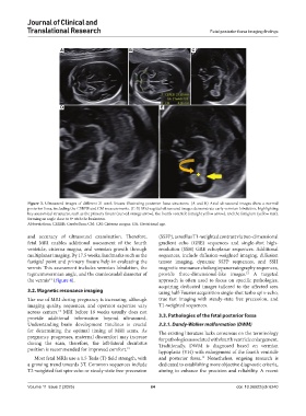
Figure 3. Ultrasound images of different 21-week fetuses illustrating posterior fossa structures. (A and B) Axial ultrasound images show a normal
posterior fossa, including the CEREB and CM measurements. (C-E) Mid-sagittal ultrasound images demonstrate early vermian lobulation, highlighting
key anatomical structures, such as the primary fissure (curved orange arrow), the fourth ventricle (straight yellow arrow), and the fastigium (yellow star),
forming an angle close to 0º with the brainstem.
Abbreviations: CEREB: Cerebellum; CM: CM: Cisterna magna; GA: Gestational age.
and accuracy of ultrasound examination. Therefore, (SSFP), as well as T1-weighted contrast via two-dimensional
fetal MRI enables additional assessment of the fourth gradient echo (GRE) sequences and single-shot high-
ventricle, cisterna magna, and vermian growth through resolution (SSH) GRE echoplanar sequences. Additional
multiplanar imaging. By 17.5 weeks, landmarks such as the sequences, include diffusion-weighted imaging, diffusion
fastigial point and primary fissure help in evaluating the tensor imaging, dynamic SSFP sequences, and SSH
vermis This assessment includes vermian lobulation, the magnetic resonance cholangiopancreatography sequences,
tegmentovermian angle, and the craniocaudal diameter of provide three-dimensional-like images. A targeted
12
the vermis (Figure 4). approach is often used to focus on specific pathologies,
11
acquiring dedicated images tailored to the affected area
3.2. Magnetic resonance imaging using half-Fourier acquisition single-shot turbo spin-echo,
The use of MRI during pregnancy is increasing, although true fast imaging with steady-state free precession, and
imaging quality, sequences, and operator expertise vary T1-weighted sequences.
across centers. MRI before 18 weeks usually does not
12
provide additional information beyond ultrasound. 3.3. Pathologies of the fetal posterior fossa
Understanding brain development timelines is crucial 3.3.1. Dandy-Walker malformation (DWM)
for determining the optimal timing of MRI scans. As The existing literature lacks consensus on the terminology
pregnancy progresses, maternal discomfort may increase for pathologies associated with fourth ventricle enlargement.
during the scan, therefore, the left-lateral decubitus Traditionally, DWM is diagnosed based on vermian
position is recommended for improved comfort. 13
hypoplasia (VH) with enlargement of the fourth ventricle
Most fetal MRIs use a 1.5 Tesla (T) field strength, with and posterior fossa. Nonetheless, ongoing research is
14
a growing trend towards 3T. Common sequences include dedicated to establishing more objective diagnostic criteria,
T2-weighted fast spin-echo or steady-state free-precession aiming to enhance the precision and reliability. A recent
Volume 11 Issue 2 (2025) 64 doi: 10.36922/jctr.6240