
Page 25 - JCTR-9-4
P. 25
Marchand et al. | Journal of Clinical and Translational Research 2023; 9(4): 236-245 241
therapy, which included two or three doses of 2–4 mL of the (RR = 0.78, 95% CI [0.50, 1.20], P = 0.26), as shown in Figure 6.
20% fat emulsion each diluted in saline, given in the time period We found homogeneity among the pooled studies (P = 0.38,
surrounding embryo transfer [25-29]. While these protocols were I² = 0%). The quality of evidence was moderate, as shown in
similar in timing and dosage, they were not identical. Figure 4.
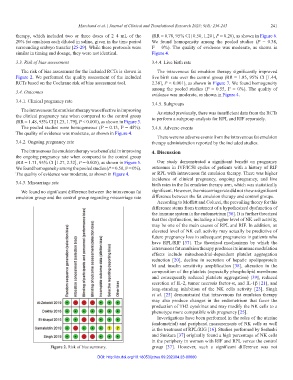
3.3. Risk of bias assessment 3.4.4. Live birth rate
The risk of bias assessment for the included RCTs is shown in The intravenous fat emulsion therapy significantly improved
Figure 2. We performed the quality assessment of the included live birth rate over the control group (RR = 1.85, 95% CI [1.44,
RCTs based on the Cochrane risk of bias assessment tool. 2.38], P < 0.001), as shown in Figure 7. We found homogeneity
among the pooled studies (P = 0.55, I² = 0%). The quality of
3.4. Outcomes
evidence was moderate, as shown in Figure 4.
3.4.1. Clinical pregnancy rate
3.4.5. Subgroups
The intravenous fat emulsiontherapy was effective in improving
As stated previously, there was insufficient data from the RCTs
the clinical pregnancy rate when compared to the control group to perform a subgroup analysis for RPL and RIF separately.
(RR = 1.48, 95% CI [1.23, 1.79], P < 0.001), as shown in Figure 3.
The pooled studies were homogeneous (P = 0.13, I² = 45%). 3.4.6. Adverse events
The quality of evidence was moderate, as shown in Figure 4.
There were no adverse events from the intravenous fat emulsion
3.4.2. Ongoing pregnancy rate therapy administration reported by the included studies.
The intravenous fat emulsion therapy was beneficial in improving 4. Discussion
the ongoing pregnancy rate when compared to the control group
(RR = 1.71, 95% CI [1.27, 2.32], P = 0.005), as shown in Figure 5. Our study demonstrated a significant benefit on pregnancy
We found homogeneity among the pooled studies (P = 0.50, I² = 0%). outcomes in IVF/ICSI cycles of patients with a history of RIF
The quality of evidence was moderate, as shown in Figure 4. or RPL with intravenous fat emulsion therapy. There was higher
incidence of clinical pregnancy, ongoing pregnancy, and live
3.4.3. Miscarriage rate birth rates in the fat emulsion therapy arm, which was statistically
We found no significant difference between the intravenous fat significant. However, the miscarriage rate did not show a significant
emulsion group and the control group regarding miscarriage rate difference between the fat emulsion therapy and control groups.
According to Moffett and Colucci, the prevailing theory for this
difference stems from treatment of a hypothesized dysfunction of
the immune system in the endometrium [36]. It is further theorized
that this dysfunction, including a higher level of NK cell activity,
may be one of the main causes of RPL and RIF. In addition, an
elevated level of NK cell activity may actually be predictive of
future pregnancy loss in subsequent pregnancies in patients who
have RPL/RIF [37]. The theorized mechanisms by which the
intravenous fat emulsion therapy produces its immune modulation
effects include mitochondrial-dependent platelet aggregation
reduction [20], decline in secretion of hepatic apolipoprotein
M and insulin sensitivity amplification [38], alteration in the
composition of the platelets (especially phospholipid membrane
and consequently reduced platelets aggregation) [39], reduced
secretion of IL-2, tumor necrosis factor-α, and IL-1β [21], and
long-standing inhibition of the NK cells activity [23]. Singh
et al. [25] demonstrated that intravenous fat emulsion therapy
may also produce changes in the endometrium that favor the
production of TH2 cytokines and may modify the NK cells to a
phenotype more compatible with pregnancy [25].
Investigations have been performed in the roles of the uterine
(endometrial) and peripheral measurements of NK cells as well
in the treatment of RPL/RIG [16]. Studies performed by Seshadri
and Sunkara [37] originally found a high percentage of NK cells
in the periphery in women with RIF and RPL versus the control
Figure 2. Risk of bias summary. group [37]. However, such a significant difference was not
DOI: http://dx.doi.org/10.18053/jctres.09.202304.23-00060