
Page 75 - JCTR-9-4
P. 75
Xu et al. | Journal of Clinical and Translational Research 2023; 9(4): 290-296 291
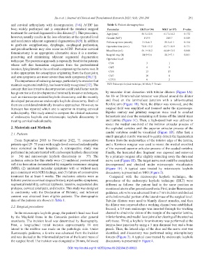
and cervical arthroplasty with decompression [3-6]. ACDF has Table 1. Patient demographics
been widely performed and is considered the standard surgical Parameter EKT (n=34) MKT (n=37) P‑value
treatment for cervical degenerative disc disease [7]. This procedure, Age (year) 56.5±12.8 61.7±14.2 0.172
however, usually results in the loss of motion at the operated level Gender (M/F) 15/19 20/17 0.549
and accelerates adjacent segmental degeneration [8,9]. In addition Follow-up time (month) 31.8±6.3 29.5±5.1 0.154
to graft-site complications, dysphagia, esophageal perforation, Operative time (min) 71.0±15.2 63.7±18.9 0.131
and pseudoarthrosis may also occur in ACDF. Posterior cervical blood loss (ml) 56.1±18.2 64.4±13.5 0.068
foraminotomy is an appropriate alternative since it is a motion- hospital stay (h) 24.9±5.6 28.3±7.1 0.061
preserving and minimizing adjacent segmental degeneration Operative level
technique. The posterior approach is especially feasible for patients
whose soft disc herniation originates from the posterolateral C3/4 3 1
location, lying lateral to the cord and compressing the nerve root. It C4/5 8 9
is also appropriate for osteophytes originating from the facet joint, C5/6 13 17
and arm symptoms are more severe than neck symptoms [10,11]. C6/7 10 8
The importance of reducing damage, particularly to muscles that C7/T1 0 2
maintain segmental stability, has been widely recognized [12]. The EKT: Endoscopic keyhole technique; M: Male; F: Female
concept that less invasive decompression could yield better results
has given rise to the development of minimally invasive techniques, by muscular blunt dissection with tubular dilators (Figure 1A).
such as microscope-assisted keyhole discectomy and the recently An 18- or 20-mm tubular retractor was placed around the dilator
developed percutaneous endoscopic keyhole discectomy. Both of and fixed on the laminofacet junction with a table-mounted
them are considered minimally invasive approaches. However, no flexible arm (Figure 1B). Next, the dilator was removed, and the
literature has reported which one is superior in treating cervical surgical field was amplified and focused under the microscope.
radiculopathy. This study aimed to compare the clinical outcomes Bipolar cautery and pituitary rongeurs were used to conduct
of endoscopic keyhole and microscopic keyhole discectomy in hemostasis and clear the remaining soft tissue off the lateral mass
treating cervical radiculopathy. and lamina (Figure 1C). Then, a high-speed burr was utilized to
resect the medial one-third of the inferior articular process of
2. Materials and Methods the cephalad vertebra until the superior articular process of the
2.1. Patients caudal vertebrae could be visualized (Figure 1D). After that, a
small upangled curette was used to gently detach the ligamentum
From September 2018 to November 2022, 71 consecutive flavum from the undersurface of the inferior edge of the lamina,
patients aged 29–75 years with single-level cervical radiculopathy and a Kerrison rongeur was used to resect the medial one-third
were reviewed in four hospitals. A retrospective study was of the exposed superior articular process of the caudad vertebra.
performed in patients treated with endoscopic keyhole discectomy Finally, the herniated disc fragment was exposed and removed
(n = 34) and microscopic keyhole discectomy (n = 37). The by a pituitary rongeur after slightly retracting away the dura and
inclusion criteria for this study were (1) unilateral posterolateral nerve root (Figure 1E). The target nerve root could be completely
soft disc herniation demonstrated by magnetic resonance imaging decompressed and checked under microscopic visualization
(MRI), (2) unilateral radicular symptoms with or without neck (Figure 1F). A typical case treated by microscopic keyhole
pain consistent with MRI findings, and (3) failure of conservative discectomy is presented on MRI (Figure 2).
treatment for at least 6 weeks. The exclusion criteria were as Compared with the microscopic keyhole technique, the
follows: previous cervical surgical history, myelopathic symptoms, procedures of the endoscopic keyhole technique (EKT) were
segmental instability, cervical kyphosis, massive, sequestered disc different as follows: the patient laid in the same position as
prolapse, cervical axial pain, and discitis. This study was designed mentioned above after general anesthesia. First, under fluoroscopic
in conformity with the Declaration of Helsinki, and informed guidance, a K-wire was advanced from a 7 mm incision and docked
consent was obtained from eligible patients. The demographic at the inferomedial portion of the lateral mass of the surgical level.
data of the patients are shown in Table 1. Tubular dilators were used to bluntly dissect muscles, and then
the dilator was removed after a working channel was established.
2.2. Surgical procedures
Second, a 5.9 mm endoscope was inserted through the working
In the microscopic keyhole group, the patient’s head was fixed channel to obtain the vision of the margin of the superior lamina,
by the Mayfield frame in the Concorde position after general inferior lamina, and medial facet joint after clearing off the attached
anesthesia. The incision level was determined by fluorography. soft tissue. Third, a keyhole foraminotomy was performed at the
First, a longitudinal initial incision approximately 10 mm lamina-facet junction by using a 3 mm diamond burr and a bone
lateral to the midline was made on the pathologic side. Under punch. Then, the lateral edge of the dura and the nerve root was
fluoroscopic guidance, a K-wire was advanced from the incision identified, and discectomy was performed using micropituitary
and was docked at the inferomedial portion of the lateral mass of forceps (Figure 3). A typical case treated by endoscopic keyhole
the surgical level. The incision was elongated to 20 mm, followed discectomy is presented in Figure 4.
DOI: http://dx.doi.org/10.18053/jctres.09.202304.23-00023