
Page 77 - JCTR-9-4
P. 77
Xu et al. | Journal of Clinical and Translational Research 2023; 9(4): 290-296 293
A B
C
D
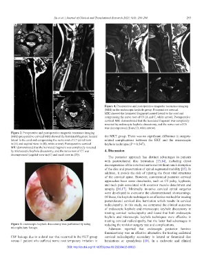
Figure 4. Preoperative and postoperative magnetic resonance imaging
(MRI) in the endoscopic keyhole group. Preoperative cervical
MRI showed the herniated fragment located lateral to the cord and
compressing the nerve root of C6 (A and C, white arrow). Postoperative
cervical MRI demonstrated that the herniated fragment was completely
resected by endoscopic keyhole discectomy, and the nerve root of C6
was decompressed (B and D, white arrow).
Figure 2. Preoperative and postoperative magnetic resonance imaging
(MRI) preoperative cervical MRI showed the herniated fragment located the MKT group. There was no significant difference in surgery-
lateral to the cord and compressing the nerve root of C7 (axial view related complications between the EKT and the microscopic
in (A) and sagittal view in (B), white arrow). Postoperative cervical keyhole technique (P = 0.547).
MRI demonstrated that the herniated fragment was completely resected
by microscopic keyhole discectomy, and the nerve root of C7 was 4. Discussion
decompressed (sagittal view in (C) and axial view in (D)).
The posterior approach has distinct advantages in patients
with posterolateral disc herniation [13,14], including direct
decompression of the involved nerve root without much disruption
of the disc and preservation of spinal segmental mobility [15]. In
addition, it avoids the risk of injuring the front vital structures
of the cervical spine. However, conventional posterior cervical
approaches have some drawbacks, such as C5 palsy, kyphosis,
and neck pain associated with extensor muscle detachment and
atrophy [16,17]. Minimally invasive cervical spinal surgeries
were developed to overcome the aforementioned shortcomings.
Of those, the keyhole technique is an effective method for treating
posterolateral cervical disc herniation which results in cervical
radiculopathy. In this study, we compared the clinical outcomes
of endoscopic keyhole and microscopic keyhole discectomy in
treating cervical radiculopathy and found that both endoscopic
keyhole and microscopic keyhole techniques were effective in
treating cervical radiculopathy, but the latter had advantages in
Figure 3. Endoscopic keyhole discectomy was performed by using reducing the revision surgery rate and complications.
micropituitary forceps. Adamson reported that endoscopic posterior lamino-
foraminotomy was an effective alternative for treating unilateral
CSF leakage due to a dural tear that occurred in the EKT group cervical radiculopathy secondary to lateral or foraminal disc
versus 1 patient who suffered nerve root temporary irritation in herniations or spondylosis [18]. In a cadaveric and clinical
DOI: http://dx.doi.org/10.18053/jctres.09.202304.23-00023