
Page 78 - JCTR-9-4
P. 78
294 Xu et al. | Journal of Clinical and Translational Research 2023; 9(4): 290-296
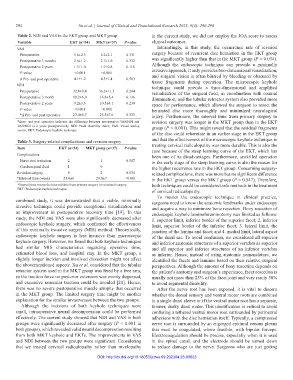
Table 2. NDI and VAS in the EKT group and MKT group in the current study, we did not employ the JOA score to assess
Variable EKT (n=34) MKT (n=37) P‑value clinical outcomes.
VAS Interestingly, in this study, the occurrence rate of revision
Preoperative 5.6±2.3 6.2±2.1 0.331 surgery because of recurrent disc herniation in the EKT group
Postoperative 3 months 2.4±1.2 2.7±1.0 0.332 was significantly higher than that in the MKT group (P = 0.034).
Postoperative 2 years 1.5±1.0 1.9±0.8 0.118 Although the endoscopic technique can provide a minimally
P-value <0.001 <0.001 invasive approach, it only provides two-dimensional visualization,
Δ Pre- and post operative 4.1±1.2 4.3±1.4 0.583 and surgical vision is often blurred by bleeding or obscured by
tissue fragments during operation. The microscopic keyhole
NDI technique could provide a three-dimensional and amplified
Preoperative 32.8±9.4 36.2±11.3 0.244 visualization of the surgical field, in coordination with coaxial
Postoperative 3 month 19.2±6.0 16.8±5.4 0.136 illumination, and the tubular retractor system also provided more
Postoperative 2 years 9.2±3.6 10.5±4.1 0.230 space for performance, which allowed the surgeon to resect the
P-value <0.001 <0.001 herniated disc more thoroughly and minimized neurological
*Δ Pre- and post operative 23.4±5.7 25.3±7.6 0.313 injury. Furthermore, the interval time from primary surgery to
*Δpre- and post operative indicates the difference between preoperative VAS/NDI and revision surgery was longer in the MKT group than in the EKT
VAS/NDI at 2 years postoperatively. NDI: Neck disability index; VAS: Visual analog group (P < 0.001). This might reveal that the residual fragments
scores; EKT: Endoscopic keyhole technique
of the disc could reherniate in an earlier stage in the EKT group
Table 3. Surgery-related complications and revision surgery and that the effectiveness of the microscopic keyhole technique in
Item EKT (n=34) MKT group (n=37) P‑value treating cervical radiculopathy was more durable. This is also the
case because of the steep learning curve of the EKT, which has
Complications been one of its disadvantages. Furthermore, unskilled operation
Nerve root irritation 2 1 0.547 in the early stage of the steep learning curve is also the reason for
Cerebrospinal fluid 1 0 the higher recurrence rate in the EKT group. Concerning surgery-
Revision surgery 9 2 0.034 related complications, there was more but no significant difference
*Interval time (week) 21.0±6 29.0±7 <0.001 in the EKT group versus the MKT group (P = 0.547). Therefore,
*Interval time means the interval time from primary surgery to revisional surgery. both techniques could be considered safe methods in the treatment
EKT: Endoscopic keyhole technique
of cervical radiculopathy.
To master the endoscopic technique in clinical practice,
combined study, it was demonstrated that a viable, minimally surgeons need to know the anatomic landmarks under endoscopy
invasive technique could provide exceptional visualization and and acquire a way to minimize bone resection. Bony resection of
an improvement in postoperative recovery time [19]. In this endoscopic keyhole laminoforaminotomy was limited as follows:
study, the NDI and VAS were also significantly decreased after 1. superior limit, inferior border of the superior facet; 2. inferior
endoscopic keyhole surgery, which confirmed the effectiveness limit, superior border of the inferior facet; 3. lateral limit, the
of this minimally invasive surgery (MIS) method. Theoretically, junction of the lamina and facet; and 4. medial limit, lateral aspect
endoscopic keyhole surgery is less invasive than microscopic of the dural sac. To avoid confusion, we considered all superior
keyhole surgery. However, we found that both keyhole techniques and inferior anatomic structures of a superior vertebra as superior
had similar MIS characteristics regarding operative time, and all superior and inferior structures of an inferior vertebra
estimated blood loss, and hospital stay. In the MKT group, a as inferior. Hence, instead of using anatomic nomenclature, we
slightly longer incision and involved dissection might not affect identified the facets and laminae based on their relative surgical
the abovementioned aspects. Xu et al. considered that the tubular perspectives. Although the amount of bony resection depends on
retractor system used in the MKT group was fixed by a free arm, the patient’s anatomy and surgeon’s experience, facet resection is
so the traction force on posterior extensors was evenly dispersed, usually not more than 25% of the facet joint and very rarely 50%
and excessive muscular traction could be avoided [20]. Hence, to avoid segmental disability.
there was no severe postoperative muscle atrophy that occurred After the nerve root has been exposed, it is vital to discern
in the MKT group. The limited surgery time might be another whether the dorsal sensory and ventral motor roots are combined
explanation for the similar invasiveness between the two groups. in a single dural sleeve or if the ventral motor root has a separate,
Although the incisions of both keyhole techniques were thinner, dusky dural mater. This identification is critical to avoid
small, intraoperative neural decompression could be performed confusing a tethered ventral motor root surrounded by perineural
effectively. The current study showed that NDI and VAS in both adhesions with the disc herniation itself. Typically, a compressed
groups were significantly decreased after surgery (P < 0.001 in nerve root is surrounded by an engorged epidural venous plexus
both groups), which revealed valid neural decompression resulting that must be coagulated, where feasible, with bipolar forceps.
from both MKT keyhole and EKTs. The improvements in VAS Electrocoagulation should be precise, especially when it is used
and NDI between the two groups were significant. Considering in the spinal canal, and the electrode should be turned down
that we treated cervical radiculopathy rather than myelopathy to reduce damage to the nerve. Surgeons who are just getting
DOI: http://dx.doi.org/10.18053/jctres.09.202304.23-00023