
Page 76 - JCTR-9-4
P. 76
292 Xu et al. | Journal of Clinical and Translational Research 2023; 9(4): 290-296
A B C
D E F
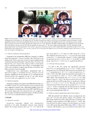
Figure 1. The procedure of the microscopic keyhole technique. (A) Lateral fluoroscopic image confirmed the interest level and demonstrated the
starting dilator advancement over the target level. (B) The final 20-mm tubular retractor was placed over the dilators and fixed into place over the
laminofacet junction with a table-mounted flexible retractor arm. (C) The surgical field was amplified and focused under the microscope, and the
inferior articular process of C6 and the superior articular process of C7 were visualized. (D) After a high-speed burr was utilized to resect the medial
third of the inferior articular process of C6 and the superior articular process C7, the yellow ligament was presented. (E) After detachment of the
ligamentum flavum from the undersurface of the inferior edge of the lamina and resection of the medial third of the exposed facet joint of C6-C7, the
herniated disc and compressed C7 nerve root were exposed. (F) The herniated disc fragment was removed by a pituitary rongeur, and then the C7 nerve
root was completely decompressed.
2.3. Clinical evaluations and management EKT group and 63.7 ± 18.9 min in the MKT group (P = 0.131).
The estimated blood loss was 56.1 ± 18.2 ml in the EKT group
Preoperative and postoperative MRI was compared to evaluate versus 64.4 ± 13.5 ml in the MKT group (P = 0.068). Additionally,
neural decompression. The neck disability index (NDI) and visual the hospital stay (24.9 ± 5.6 h vs. 28.3 ± 7.1 h for EKT vs. MKT
analog scale (VAS) scores were recorded to assess intragroup and group, respectively, P = 0.061) was not significantly different.
intergroup neurological functions. Operative time, blood loss, and
hospital stay were documented. Surgery-related complications 3.2. NDI and VAS assessments
such as neurological deficits and leakage of cerebrospinal fluid
(CSF) were recorded to evaluate surgical safety. All patients were The NDI in the EKT group was significantly decreased
followed up for at least 24 months. from 32.8 ± 9.4 preoperatively to 9.2 ± 3.6 (P < 0.001) 2 years
Antimicrobials were intravenously administered half an postoperatively. The NDI in the MKT group decreased from 36.2
hour before surgery in all patients just once. Analgetic acid was ± 11.3 preoperatively to 10.5 ± 4.1 2 years postoperatively (P <
routinely administered for all patients for 72 h postoperatively. 0.001). VAS in the EKT group decreased from preoperative 5.6
General activity was suggested on the 2 day after surgery. ± 2.3 to postoperative 2 years 1.5 ± 1.0 (P < 0.001), while in the
nd
A cervical collar was suggested for use for 2 weeks. MKT group, VAS decreased from 6.2 ± 2.1 to 1.9 ± 0.8 after
surgery 2 years (P < 0.001). The improvement in NDI in the EKT
2.4. Statistical analysis group and that in the MKT group were not significantly different
(23.4 ± 5.7 vs. 25.3 ± 7.6, P = 0.313). The same was true for the
All statistical analyses were performed by IBM SPSS Statistics ver.
19.0 (IBM, Armonk, NY, USA). Preoperative and postoperative data improvements in VAS between the two groups (4.1 ± 1.2 vs. 4.3
were compared by paired t-tests. Independent samples t-tests were ± 1.4, P = 0.583). Comparing with the EKT group, the VAS and
used to compare corresponding data between EKT and MKT groups. NDI were similarly ameliorated in the MKT group at 3 months
The revision surgery rate and complication rate were compared by and 2 years postoperatively (Table 2).
the Chi-square test. Data are presented as mean ± standard deviation. 3.3. Surgery-related complications and revision surgery
A P ≤ 0.05 was considered statistically significant.
MRI demonstrated that effective neural decompression was
3. Results observed in all cases after primary surgery. Nine patients in the
EKT group underwent revision surgery because of recurrent disc
3.1. Operative outcomes
herniation versus 2 patients in the MKT group (P = 0.034). The
Seventy-one consecutive patients were retrospectively interval time from primary surgery to revision surgery was shorter
reviewed in this study. All patients’ incisions were primarily in the EKT group than in the MKT group (Table 3). There were
healed. The average operative time was 71.0 ± 15.2 min in the 2 patients with temporary nerve root irritation and 1 patient with
DOI: http://dx.doi.org/10.18053/jctres.09.202304.23-00023