
Page 78 - MI-1-1
P. 78
Microbes & Immunity An ATP-free packaging of T4 DNA
A C Pi and Ca could determine the conformation of DNA,
2+
which triggers either the packaging or the ejection of
DNA. Specifically, increasing the concentration of Ca
2+
or decreasing the Pi concentration could induce the
packaging of DNA (Table 1).
The loss of infectivity of condensed DNA without
capsid was confirmed with proteinase K treatment of the
virions. The capsid protein of virions was degraded by
proteinase K (Figure 2B). The subsequent on-filter-dry
B D DNase I treatment degraded all the virion-like particles
(Figure 2C), leaving behind naked DNA particles. The
treatment of inactivated proteinase K also produced
similar virion-like particles, which were resistant to DNase
I degradation (Figure 2D). The infectivity, measured
by pfu, of the suspensions treated with proteinase K
was <1/10,000 that of the T4 populations treated with
inactivated proteinase K. Globular naked T4 DNA was
unable to infect E. coli cells. This also confirms that the
infectious bodies regenerated through the adjustment of
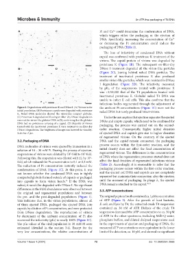
Figure 2. Degradations with proteinase K and DNase I. (A) Virions in the the ambient Pi concentrations (Figure 1C) were not the
initial population. (B) Proteinous capsids were degraded with proteinase naked DNA but newly produced intact virions.
K. Naked DNA molecules showed the virion-like compact globules.
(C) Proteinase K degradation following on-filter-dry DNase I degradation Under the assumption that ejection separates the ejected
removed the virion-like globular DNA in (B), confirming that the globular DNAs and empty capsids, which need to be combined for
DNA had no proteinous covering of a capsid. (D) Majority of virions packaging, the process was carried out on the second-
treated with the inactivated proteinase K were resistant to on-filter-dry
DNase I degradation. The brightness of images was inverted for visuality. order reaction. Consequently, higher initial densities
Scale bar: 5 μm. of ejected DNA and capsids give rise to higher densities
of regenerated virions. On the contrary, if the ejected
3.2. Packaging of DNA DNA and the capsid remain connected, the packaging
DNA molecules of virions were ejected by immersion in a process occurs within the first-order reaction, and the
solution of 10 – 30 mM Pi. During the process of ejection, initial density does not affect the final concentration of
suspensions of virions were diluted by 10 -fold to 10 -fold. regenerated virions. The differences in the concentrations
2
5
Following this, the suspension was diluted with EL by 10 - of DNA when the regeneration processes started does not
2
fold, which reduced the Pi concentration to 0.1 or 0.3 mM. affect the final densities of regenerated infectious virions
The reduction of Pi concentration instantly induced the (Table 2). Accordingly, it is reasonable to infer that the
condensation of DNA (Figure 1C). At this point, it was packaging process occurs within the first-order reaction,
not known whether the condensed DNA was in tightly and the ejected coil DNA and capsids are not completely
compacted globule formed outside of capsids or packaged separated but maintain their connection after the ejection
into capsids to form virion heads. If the DNA was until the moment of packaging. In phage λ, the ejected
25
naked, it would be degraded with DNase I. No significant DNA remains attached to the capsid. 26,27
differences in the FLM abundances were observed between 3.3. ATP concentrations
the original and regenerated populations and between
the pre- and the post-degraded populations (Figure 1). The original peptone broth contained ca. 1 μM concentration
This indicates that, in the virion populations, almost all of ATP (Figure 3). After the growth of host bacteria,
of them ejected DNA, packaged the ejected DNA into E. coli, and lysis by T4, the extracted crude T4 suspension
capsids by dilution of Pi concentration, and were protected contained ca. 30 nM of ATP. Dialysis of the crude T4
from DNase degradation. The reproduction of virions suspension decreased the ATP concentration to ca. 30 pM
by decrement of the ambient concentration of Pi also of ATP. In the other specimens, including Milli-Q water,
recovered the infectivity (pfu) to nearly 100% (Figure 1C). phosphate buffers, and diluted dialyzed suspensions used
The pfu values of the viral population in 30 mM Pi were in the experiments of ejection and packaging of DNA, the
estimated (detailed in the section 3.4). Except for the measured ATP concentrations were equivalent to the lower
very low concentrations, the relative concentrations of limit of the detection, ca. 10 pM, and showed no significant
Volume 1 Issue 1 (2024) 72 doi: 10.36922/mi.2666