
Page 79 - MI-1-1
P. 79
Microbes & Immunity An ATP-free packaging of T4 DNA
30 pM of ATP. This low concentration can be practically
construed as ATP-free in the context of DNA packaging.
3.4. pfu values of 30 mM Pi specimens and the
activities of DNase I in EL, 30 mM Pi and TE
The behaviors of DNase I in the conditions used in this
study (i.e., through in situ degradations and on-filter
degradations of DNA molecules versus in EL, 10 – 30 mM
Pi and TE) were elucidated (Figure 4).
In the T4 suspensions in EL, the abundances of virion-
like globular DNA and the pfu values showed no difference
between the original specimen, in situ and on-filter DNase
I-treated specimens (Figure 4A-C). In situ and on-filter-
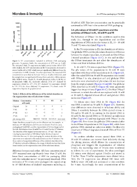
Figure 3. TP concentrations included in different DNA packaging dry DNase treatments do not affect the abundances of
processes. In peptone broth, the concentration of ATP was ca. 1 μM. virions and their infectious abilities.
The extracted crude T4 suspension included several tenths nM of ATP.
After dialysis, the ATP concentration in the T4 suspension decreased to In 30 mM Pi, T4 virions eject their DNA (Figure 4D).
several tenths pM of ATP, which was not significantly different from the The pfu values of the populations in 30 mM Pi were
concentration in Milli-Q water. The other specimens, the measured ATP equivalent with those of the populations in EL (Figure 4D).
concentrations were close to the lower limit, ca. 10 pM, of detection, and After the naked DNA in 30 mM Pi suspension was treated
the samples bore no significant difference from each other. Abbreviations: with DNase I in situ, abundant coil and globular DNA
MQ: Milli-Q water; 30mM Pi, 30 mM phosphate buffer; 0.1M Pi, 0.1
M phosphate buffer; EL, electrolyte solution; DT4 10 , dialyzed T4 molecules were observed and pfu values did not decrease
-6
suspension diluted to 10 ; DT4 10 , dialyzed T4 suspension diluted from the original sample (Figures 1A and 4E). The globular
-2
-6
to 10 ; DT4 dir, direct dialyzed T4 suspension; T4, eluted crude T4 DNA observed in 30 mM Pi (Figure 4E) were apparently
-2
suspension; Peptone br., peptone broth. bigger than intact virions (Figure 4A-C). On-filter DNase I
treatment, in which the solvent was replaced with 10 mM
Table 2. Effects of the differences of the initial densities on or 30 mM Pi, digested almost all coil and globular DNA
the regeneration rates of infectious virions
molecules (Figure 4F).
Dilution Concentration I II III IV T4 virions eject their DNA in TE (Figure 4G) the
of Pi (mM) way DNA is ejected in 30 mM Pi (Figure 4D). However,
Original 30 0 0 0 0 clear differences were observed in infectious ability and
1 dil. 30 0 −1 −2 −3 DNase I sensitivity between the DNA-ejected virions in
st
2 dil. 0.3 −2 −2 −2 −2 TE and in 30 mM Pi. In contrast to the ejected DNA in
nd
3 dil. 0.3 −3 −2 −1 0 30 mM Pi, the ejected DNA in TE showed no infectious
rd
pfu average (SD) 100.0 (15.3) 73.6 (1.0) 124.3 (11.5) 184.5 (18.2) ability (Figure 4G) and was degraded with DNase I in situ
Notes: Numbers, except for pfu, are the dilution rate, for example, -n (Figure 4H). Few virion-like globular DNA remained after
indicates 10 -fold dilution and 0 means no dilution. In all the cases, in situ DNase I degradation in TE (Figure 4H), while these
n
packaging of viral DNA, and the regeneration of virions, happened at globular DNA showed no infectious ability. On-filter-dry
the 100-fold dilution from 30 mM Pi to 0.3 mM Pi at the 2 dilution. treatment of DNase I degrades almost all DNA of T4 in TE
nd
The densities of the original specimens were ca. 10 mL . The case (Figure 4I).
-1
7
I packaging was started at 1000 times higher density of ejected
DNA than the case IV. After packaging, the content densities of the To confirm whether virions ejected their DNA in
specimens were adjusted to the same densities at the 3 dilution. The 30 mM Pi solution can recover their infectivity during
rd
three orders of magnitude difference of the substrate densities at the the plating process, which dilutes the concentration of
2 dilution induced a difference, the range of which was less than three
nd
times weaker, on the regeneration rates of the infectious virions. phosphate and triggers the re-production of infective
virions, the recovering rates of virions were monitored
difference between each other. All these concentrations of as follows. In a set of experiments, the ultra-centrifuged
ATP measured were significantly lower than 25 μM, the T4 virions were dialyzed against T-buffer and stored in
lower limit concentration of ATP for packaging DNA T-buffer. These T-buffer suspensions were diluted twice.
with the molecular motor. As previously described, DNA First, the T4 suspension was diluted 100 times with
5
molecules of T4 virions were packaged into capsids at up Milli-Q water 100-fold and acclimated to this condition
to 10 -fold dilution of dialyzed suspension including ca. for 5 min. Accordingly, the ionic concentrations of the
7
Volume 1 Issue 1 (2024) 73 doi: 10.36922/mi.2666