
Page 81 - MI-1-1
P. 81
Microbes & Immunity An ATP-free packaging of T4 DNA
The step-by-step mechanism of the ejection of DNA
was not investigated in this study. However, the completed
head and tail have been shown to join spontaneously
after DNA packaging is completed in the assembly of
bacteriophage. 32,33 Therefore, it is presumed that, in the
ejection of DNA from a capsid, the tail may detach from
the connector first and the ejection of DNA from the
connector follows, according to the increment of the
ambient phosphate concentration.
4.2. Packaging of DNA
Before packaging, it would be appropriate to examine again
if there were any intact virions in the 30 mM Pi suspension.
The FLM observations clearly indicated ejection of the
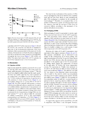
Figure 5. The time courses of pfu of T-buffer suspension (Tb), 100- and majority of DNA and almost no intact virions in 30 mM Pi
10000-fold dilutions of T-buffer suspensions (0.01Tb, 0.0001Tb), 100-
fold dilution of 0.01Tb in 30 mM phosphate buffer (pH = 7.6, ca. 60 mM (Figures 1B and 4D). After 25 mM phosphate treatment, virions
8
Na ) (30P), and 100-fold dilution of 0.01Tb in 60 mM NaCl (60Na). separate into DNA, empty heads, and tails. It has been shown
+
that almost no intact virions remained after ultracentrifuging
9
equivalent with 0.01 T-buffer suspension (Figure 5, 60 mN virions that had been chelated with of 3 mM sodium citrate.
+
Na 0.01Tb). The cation Na was only able to maintain the Thus, it is feasible to deduce that a small amount of intact
+
infectivity of the pre-incubation suspension, while other virions may remain in 30 mM Pi suspension of T4.
attributes of the suspension failed to be preserved. On the However, pfu values of 30 mM Pi suspensions were
contrary, the incubation in 30 mM Pi produces higher pfu equivalent to the original specimens (Figure 4A and D).
than the pre-incubation or the incubation in 60 mM NaCl This apparent contradiction can be attributed to the
throughout the time course (Figure 5, 30P, 0.01Tb, and change of the concentration of Pi during plating. The
60Na). The rates of pfu values of 30 mM Pi incubation to high concentrations of Pi of the T4 inocula, where virions
60 mM Na incubation was 1.57±0.18 (mean±SD). ejected their DNA, decrease when the suspensions were
+
inoculated in the peptone agar of low Pi concentration
4. Discussion for plating, which triggers the regeneration of virions
4.1. Ejection of DNA in the agar plate. To validate the virion regeneration
process, T4 suspensions in T-buffer were incubated in
High pressure inside the capsids is considered a force used
for ejecting DNA from capsids. 28,29 The stress inside the 30 mM Pi, 60 mM NaCl and Milli-Q water, and the pfu
phage capsid decreases by polyvalent cations. 30,31 The full values were monitored (Figure 5). The solution of 60 mM
Na was chosen as the equivalent cation concentration
+
capsid has a slightly smaller radius than the empty capsid. with 30 mM Pi. The infective ability was immediately
7
The latter two results imply that the internal pressure of lost when 0.01 T-buffer suspension was diluted 100-fold
the mature head is not higher than the external pressure.
The T4 DNA produced by degradation of capsid proteins into Milli-Q water (Figure 5, 0.0001Tb). The capability of
+
with proteinase K shows compact forms (Figure 2), not the monovalent cation, 60 mM Na , in preserving pfu is
self-repulsive strings like the ejected DNA in 30 mM Pi inferior to that in conserving infectious ability of the 0.01
T-buffer incubation throughout the time course (Figure 5,
(Figure 1B). The DNA molecule itself assumes a compact
conformation within and without a capsid under the regular 60P, Na, 0.01Tb). The infectivity recovery of the incubation
extracellular ambient concentrations of ions, pressure, and from 30 mM Pi, in contrast, was higher than the 0.01
temperature without addition of any multivalent cations. T-buffer inocula, and moreover, it was equivalent or higher
The globule-to-coil phase transition of phage DNA, which than the original 1 T-buffer suspension (Figure 5, 30P, Tb).
Plating of virions of which DNA was ejected in 30 mM Pi
is necessary for the transportation of phage DNA into the
host bacteria, 8,9,27-29 can be induced solely by increment triggers DNA packaging and produce infective virions on
of ambient concentration of Pi to the concentration the normal peptone agar. This process indicates not only
corresponding to cell electrolyte in T4 DNA (Figure 1). It recovering of the infective ability but also resetting some
is implicated that this process of DNA ejection may have virions contributes to the loss the infectivity of virions.
contribution to the process of DNA ejection of T4 into the To support this, digestion of the naked DNA in 30 mM
host cell of E. coli. Pi suspension with DNase I was attempted. However,
Volume 1 Issue 1 (2024) 75 doi: 10.36922/mi.2666