
Page 74 - MI-2-2
P. 74
Microbes & Immunity Biological activity of Amazonian plants
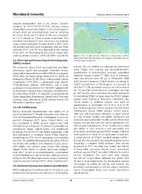
Amapari municipality) and in the Amapa “cerrado”
(savanna) at 0°55’51”N/51°11’35”W (Ferreira Gomes
municipality), respectively (Figure 1). One hundred grams
of each dried and pulverized plant material, including
the leaves of LM and the bark of OH, were macerated
in 1 L of ethanol for 7 days at room temperature. This
procedure was repeated 3 times. The extract was obtained
using a rotary evaporator (Quimis, Brasil) at 40°C under
low pressure and kept under refrigeration until use. Plant
materials of OH and LM were deposited at the Institute
of Scientific and Technological Research of Amapa State
under registration numbers 16593 and 16594, respectively. Figure 1. Sites of plant material collection in Amapa state, northern
Brazil. Notes: Black pin: Pedra Branca do Amapari municipality; Red pin:
2.2. Ultra-high-performance liquid chromatography Ferreira Gomes municipality.
(UHPLC) analysis
The methanolic extract of LM was analyzed by ultra-high- extracts. The cell viability was subsequently determined
performance liquid chromatography (Shimadzu Nexera, using Trypan blue staining and spectrophotometric
Japan), which composed of a controller (CBM 20-A), a degasser quantification of mitochondrial succinate tetrazolium
(DGU-20A), two binary pumps (Nexera X2 LC-30AD), an reductase enzyme activity. 29,30 After 24 h of treatment,
autoinjector (Nexera X2 SIL-30AC), a thermostable column cells were infected with 100 µL of SIVmac251 (NIH
compartment (CTO-20AC), a photodiode array detector AIDS Research Reagents, United States), containing an
(PDA)/ultraviolet detector (SPD-M20A), and a triple SIVp27 concentration of 599.64 pg/mL, as a measure of
27
quadrupole mass spectrometer (LC-MS-8030) equipped with viral load. Cell supernatant samples were harvested at
an electrospray ionization source. Separation was achieved on 48, 144, and 240 h post-infection, centrifuged, and kept
an LC18 column of 100 × 2.1 mm and 1.6 µm particle size at −20°C for later use in antiretroviral activity assessment
(Luna Omega Polar, Phenomenex, United States). Data were by quantifying SIVp27 antigen using the SIVp27 antigen
processed using LabSolutions LCMS software version 5.96 capture assay (Advanced Bioscience Laboratory, Inc,
(Shimadzu Corporation, Japan). 13,15 United States). In addition, samples were used for
quantification of interleukin (IL)-4, IL-6, IL-8, IL-10,
2.3. Cell viability assay and interferon-gamma (IFN-γ) using the DuoSet ELISA
Human leukocytes transformation was carried out as Development System (R&D Systems, United States).
described elsewhere. 26,27 Briefly, leukocytes were obtained After each cell supernatant collection, an equal amount
from total heparinized blood by centrifugation in a sucrose of 3 mL of fresh medium was added. SIVmac251 was
gradient (Histopaque-1077, Sigma, United States), and previously produced in Hut-78 cells, and the SIVp27viral
then maintained in RPMI medium supplemented with antigen was purified from the culture supernatant. For
10% fetal bovine serum and 1% antibiotics (penicillin and viral antigen quantification, 25 µL of disruption buffer
streptomycin, Sigma, United States), and transformed was added to each microtiter well pre-coated with
by adding 10% of Hut-78 cell culture supernatant. Cells monoclonal antibodies against SIVp27 antigen, followed
28
were cultivated in an incubator (Sanyo, Fisher Scientific, by the addition of 100 µL of cell supernatant to each well.
United States) at 37°C, 5% CO atmosphere, and controlled A serial dilution of SIVp27 standard antigen (2,000 –
2
humidity. The established human leukocyte culture (EHLC) 62.5 pg/mL) was prepared, including the negative control
was treated with multiple serial dilutions of each extract. containing a complete RPMI medium. After 60-min
Cell viability was assessed using Trypan blue staining incubation at 37°C, the wells were washed and 100 µL
(Sigma, United States) to identify and count damaged/ of horseradish peroxidase-labeled mouse monoclonal
dead cells, as well as spectrophotometric measurement antibody against SIVp27 was added, followed by another
29
of mitochondrial succinate tetrazolium reductase enzyme 60-min incubation. The wells were then washed,
activity in the cell culture supernatant, using a commercial and 100 µL of peroxidase substrate was added. After
cytotoxic assay kit (CytoSkan WST-1, Roche, Switzerland). 30 30 min at room temperature, the chromogenic reaction
was stopped by adding 2N sulfuric acid solution. The
2.4. Antiretroviral assay and cytokine quantification absorbance was measured at 450 nm using a microplate
EHLC, cultivated in 25 cm flasks with 5 × 10 cells/mL, was reader (Thermoplate, Italy), with results expressed in
2
5
treated with a low cytotoxic concentration (1:1024) of plant optical density.
Volume 2 Issue 2 (2025) 66 doi: 10.36922/mi.8367