
Page 76 - MI-2-2
P. 76
Microbes & Immunity Biological activity of Amazonian plants
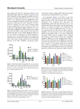
and infected with SIV, IL-4 expression (Figure 4) was treated cells to extract-treated and SIV- infected cells, there
significantly higher at the 48-h interval in comparison to all were significant statistical differences (p=0.0040).
other treatments (p-value ranging from 0.0021 to 0.0210), IL-6 expression (Figure 5) by EHLC at the 48-h
except in LM extract treatment (p=0.1057). At the 144-h interval showed a significant difference (p=0.0006), with
interval, IL-4 expression remained significantly higher higher secretion in LM extract-treated cells. Comparing
(p-value ranging from 0.0004 to 0.0022) than in all other SIV-infected cells, including those previously and
treatments, including LM extract treatment combined independently treated with LM and OH extracts, it was
with SIV infection and LM extract treatment alone observed that OH extract-treated cells had significantly
(p-value ranging from 0.2736 to 0.3056). At 240 h, IL-4 higher IL-6 secretion (p=0.0030). Cells treated with LM
expression was significantly higher compared to all other extract alone produced significantly more IL-6 than in
treatments, including SIV infection alone (p-value ranging OH extract-treated and SIV-infected cells (p=0.0033).
from 0.0197 to 0.0205). Cells treated with LM extract and Similarly, LM extract-treated cells had significantly
infected with SIV did not display significant differences higher IL-6 production than in OH extract-treated cells
in IL-4 expression compared to all other treatments at (p=0.0049). Other treatment conditions, with or without
any time interval. In the initial stage of the experiment, SIV infection, as well as controls, did not show statistically
differences were not significant between cells treated with significant differences (p-value ranging from 0.0001 to
either extract alone or when infected by SIV (p>0.9999). 0.1467). At the 144-h interval, the highest IL-6 secretion
However, in all other conditions, comparing extract-
(p<0.0001) was observed in cells treated with OH extract
alone compared to LM extract-treated and SIV-infected
cells, as well as in comparison to OH extract-treated and
SIV-infected cells and controls (p=0.011). No statistically
significant differences were observed in other conditions.
Figure 2. Growth of the established human leukocyte cell line population
under treatment with Licania macrophylla and Ouratea hexasperma
extracts over a 288-h culture period. At the initial time point of 0 h,
all cells, both treated and untreated had the same population density. Figure 4. Interleukin-4 expression in an established human leukocyte cell
Note: *indicates significant at p<0.05. line treated with Licania macrophylla and Ouratea hexasperma, followed
by SIV infection, and in control cells treated with extracts but not infected
with SIV. Note: *indicates significant at p<0.05.
Abbreviation: SIV: Simian immunodeficiency virus.
Figure 3. Established human leukocyte cell line treated for 24 h with
Licania macrophylla and Ouratea hexasperma extracts, independently, Figure 5. Interleukin-6 expression in an established human leukocyte cell
and subsequently infected with SIVmac251. Cell viability was assessed up line treated with Licania macrophylla and Ouratea hexasperma, followed
to 288 h of culture. At the initial time point of 0 h, all cells had the same by SIV infection, and in control cells treated with extracts but not infected
population density. Note: *indicates significant at p<0.05. with SIV. Note: *indicates significant at p<0.05.
Abbreviation: SIV: Simian immunodeficiency virus. Abbreviation: Simian immunodeficiency virus.
Volume 2 Issue 2 (2025) 68 doi: 10.36922/mi.8367