
Page 121 - MI-2-4
P. 121
Microbes & Immunity Isolation and identification of PEDV strain CHN-CQ-2021
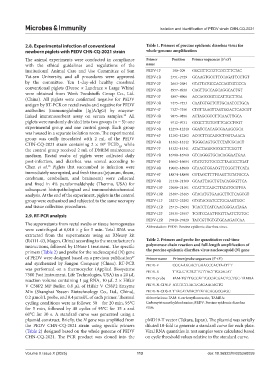
2.8. Experimental infection of conventional Table 1. Primers of porcine epidemic diarrhea virus for
newborn piglets with PEDV CHN-CQ-2021 strain whole‑genome amplification
The animal experiments were conducted in compliance Primer Position Primer sequences (5’→3’)
with the ethical guidelines and regulations of the name
Institutional Animal Care and Use Committee of Sun PEDV-1F 190–209 GGCGTTCCGTCGCCTTCTAC
Yat-sen University, and all procedures were approved PEDV-1R 2751–2729 GCAAGTGCCTTCCAGATTCCTGT
by the committee. Ten 1-day-old healthy crossbred PEDV-2F 2663–2684 GTATTATGCCACCAGTGTCCCA
conventional piglets (Duroc × Landrace × Large White) PEDV-2R 4957–4938 CAGTTGCCAGCAGGCACTGT
were obtained from Wen’s Foodstuffs Group Co., Ltd.
(China). All piglets were confirmed negative for PEDV PEDV-3F 4887–4906 ACCAGCGGTGCATTGCTTGA
antigen by RT-PCR on rectal swabs and negative for PEDV PEDV-3R 7475–7453 CAATGTGCTCTTGCAATCCTGCA
antibodies (immunoglobulin [Ig]A/IgG) by enzyme- PEDV-4F 7327–7350 CTGTTAAGTTAGTGGACTCAGCGT
linked immunosorbent assay on serum samples. All PEDV-4R 9875–9856 ACTAGCGCCTTCAACTTGCA
25
piglets were randomly divided into two groups (n = 5): one PEDV-5F 9712–9731 GCGCTTGTGGTTCACCTGGT
experimental group and one control group. Each group PEDV-5R 12259–12240 GGATCCACAGCGAAAGCGCA
was housed in a separate isolation room. The experimental PEDV-6F 12182–12202 ACGCTTGCAGGCTGGTAAACA
group was orally inoculated with 2 mL of the PEDV
CHN-CQ-2021 strain containing 2 × 10 TCID , while PEDV-6R 14462–14442 TGGGCAGTGCTCTATCGCACT
6
50
the control group received 2 mL of DMEM maintenance PEDV-7F 14322–14341 ATACTAGGGGCGCTTCGGTT
medium. Rectal swabs of piglets were collected daily PEDV-7R 16780–16760 GTCAGGGTGCACAGGAATGAA
post-infection, and diarrhea was scored according to PEDV-8F 16662–16684 GTATGTGTGCCCTTAAGCCTGAT
Chen et al. Piglets that succumbed to infection were PEDV-8R 19002–18980 GTAAGTGGACGTTCGGCTTCATA
26
immediately necropsied, and fresh tissues (jejunum, ileum, PEDV-9F 18874–18898 CGTAGCTTTTGAGTTGTATGCCA
cerebrum, cerebellum, and brainstem) were collected PEDV-9R 21330–21309 GCAATTAGCTGTACAGGGTTCA
and fixed in 4% paraformaldehyde (Thermo, USA) for
subsequent histopathological and immunohistochemical PEDV-10F 21080–21101 CCATTCCAGCTTATATGCGTGA
analysis. At the end of the experiment, piglets in the control PEDV-10R 23487–23465 GTACATGTGAAGCTTCTCAGCGT
group were euthanized and subjected to the same necropsy PEDV-11F 23272–23292 GTGTACGATCCTGCAAGTGGC
and tissue collection procedures. PEDV-11R 25715–25694 TCACCTCATCAACGGGAATAGA
2.9. RT-PCR analysis PEDV-12F 25535–25557 TCGTCCAATTGGTTAATCTGTGC
PEDV-12R 27840–27820 TACCGTTGTGTGCAAGACCAA
The supernatants from rectal swabs or tissue homogenates Abbreviation: PEDV: Porcine epidemic diarrhea virus.
were centrifuged at 6,010 × g for 5 min. Total RNA was
extracted from the supernatants using an RNeasy kit
(R4111-03, Magen, China) according to the manufacturer’s Table 2. Primers and probe for quantitative real‑time
instructions, followed by DNase I treatment. The specific polymerase chain reaction and full‑length amplification of
primers (Table 2) and probe for the nucleocapsid (N) gene the porcine epidemic diarrhea virus nucleocapsid (N) gene
of PEDV were designed based on a previous publication Primer name Primer/probe sequences (5’→3’)
27
and synthesized by Sangon Company (China). RT-PCR PEDV-F CGCAAAGACTGAACCCACTAATTT
was performed on a thermocycler (Applied Biosystems PEDV-R TTGCCTCTGTTGTTACTTGGAGAT
7500 Fast instrument, Life Technologies, USA) in a 20 μL
reaction volume containing 1 μg RNA, 10 μL 2 × Hifair PEDV-probe FAM-TGTTGCCATTGCCACGACTCCTGC-TAMRA
V C58P2 MP Buffer, 0.8 μL of Hifair V C58P2 Enzyme PEDV-N-CDS-F ATGTCTGACGCAGAAGAGTG
Mix (Shanghai Yeasen Biotechnology Co., Ltd., China), PEDV-N-CDS-R TTACATATACTTATACAGGCGAGC
0.2 μmol/L probe, and 0.4 μmol/L of each primer. Thermal Abbreviations: FAM: 6-carboxyfluorescein; TAMRA:
cycling conditions were as follows: 50℃ for 20 min, 95°C Carboxytetramethylrhodamine; PEDV: Porcine epidemic diarrhea
for 5 min, followed by 40 cycles of 95°C for 15 s and virus.
60°C for 30 s. A standard curve was generated using a
plasmid construct. Briefly, the N gene was amplified from pMD19-T vector (Takara, Japan). The plasmid was serially
the PEDV CHN-CQ-2021 strain using specific primers diluted 10-fold to generate a standard curve for each plate.
(Table 2) designed based on the whole genome of PEDV Viral RNA quantities in test samples were calculated based
CHN-CQ-2021. The PCR product was cloned into the on cycle threshold values relative to the standard curve.
Volume X Issue X (2025) 113 doi: 10.36922/MI025260059