
Page 124 - MI-2-4
P. 124
Microbes & Immunity Isolation and identification of PEDV strain CHN-CQ-2021
A B
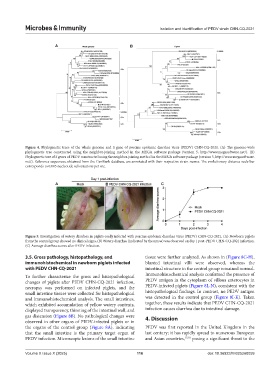
Figure 4. Phylogenetic trees of the whole genome and S gene of porcine epidemic diarrhea virus (PEDV) CHN-CQ-2021. (A) The genome-wide
phylogenetic tree constructed using the neighbor-joining method in the MEGA software package (version 5, http://www.megasoftware.net/). (B)
Phylogenetic tree of S genes of PEDV constructed using the neighbor-joining method in the MEGA software package (version 5, http://www.megasoftware.
net/). Reference sequences, obtained from the GenBank database, are annotated with their respective strain names. The evolutionary distance scale bar
corresponds to 0.005 nucleotide substitutions per site.
A B C
Figure 5. Investigation of watery diarrhea in piglets orally infected with porcine epidemic diarrhea virus (PEDV) CHN-CQ-2021. (A) Newborn piglets
from the control group showed no clinical signs. (B) Watery diarrhea (indicated by the arrow) was observed on day 1 post-PEDV CHN-CQ-2021 infection.
(C) Average diarrhea scores after PEDV infection.
3.5. Gross pathology, histopathology, and tissue were further analyzed. As shown in (Figure 8C-H),
immunohistochemical in newborn piglets infected blunted intestinal villi were observed, whereas the
with PEDV CHN-CQ-2021 intestinal structure in the control group remained normal.
To further characterize the gross and histopathological Immunohistochemical analysis confirmed the presence of
changes of piglets after PEDV CHN-CQ-2021 infection, PEDV antigen in the cytoplasm of villous enterocytes in
necropsy was performed on infected piglets, and the PEDV-infected piglets (Figure 8L-N), consistent with the
small intestine tissues were collected for histopathological histopathological findings. In contrast, no PEDV antigen
and immunohistochemical analysis. The small intestines, was detected in the control group (Figure 8I-K). Taken
which exhibited accumulation of yellow watery contents, together, these results indicate that PEDV CHN-CQ-2021
displayed transparency, thinning of the intestinal wall, and infection causes diarrhea due to intestinal damage.
gas distension (Figure 8B). No pathological changes were 4. Discussion
observed in other organs of PEDV-infected piglets or in
the organs of the control group (Figure 8A), indicating PEDV was first reported in the United Kingdom in the
that the small intestine is the primary target organ of last century; it has rapidly spread to numerous European
PEDV infection. Microscopic lesions of the small intestine and Asian countries, 17,18 posing a significant threat to the
Volume X Issue X (2025) 116 doi: 10.36922/MI025260059