
Page 17 - MSAM-2-2
P. 17
Materials Science in Additive Manufacturing Union of 2D nanomaterials and 3D printing
A B C D
E
F
H I
J
G
K
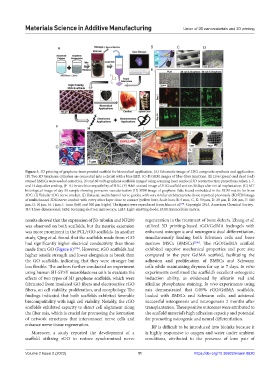
Figure 5. 3D printing of graphene-incorporated scaffold for biomedical application. (A) Schematic image of 3DG composite synthesis and application.
(B) Two 3D Graphene cylinders are connected into a circuit with a blue LED. (C–D) SEM images of fiber-fiber junctions. (E) Live (green) and dead (red)
stained hMSCs were seeded onto PLG, 20 and 60 vol% graphene scaffolds imaged using scanning laser confocal 3D reconstruction projections, taken 1, 7,
and 14 days after seeding. (F–K) In vivo biocompatibility of 3DG. (F) H&E-stained image of 3DG scaffold section 30 days after initial implantation. (G) MT
histological image of day 30 sample showing pervasive vascularization (H) SEM image of graphene flake found embedded in the ECM matrix far from
3DG. (I) Tubular 3DG nerve conduit. (J) Uniaxial, multichannel nerve guides, with very similar architectures to those reported previously. (K) SEM image
of multichannel 3DG nerve conduit with every other layer close to contact (yellow box). Scale bars: B: 5 mm, C, K: 50 μm, D: 20 μm, E: 200 μm, F: 100
μm, G: 10 μm, H: 1 μm, J: 1mm (left) and 500 μm (right). The figures were reproduced from Jakus et al. . Copyright 2015, American Chemical Society.
[94]
3D: Three-dimensional; SEM: Scanning electron microscopy; LED: Light-emitting diode; ECM: Extracellular matrix.
results showed that the expression of β3-tubulin and NF200 regeneration in the treatment of bone defects. Zhang et al.
was observed on both scaffolds, but the neurite extension utilized 3D printing-based rGO/GelMA hydrogels with
was more prominent in the PCL/rGO scaffolds. In another enhanced osteogenic and neurogenic dual differentiation,
study, Qing et al. found that the scaffolds made from rGO simultaneously loading both Schwann cells and bone
had significantly higher electrical conductivity than those marrow MSCs (BMSCs) [106] . The rGO/GelMA scaffold
made from GO (Figure 6) [105] . However, rGO scaffolds had exhibited superior mechanical properties and pore size
higher tensile strength and lower elongation at break than compared to the pure GelMA scaffold, facilitating the
the GO scaffolds, indicating that they were stronger but adhesion and proliferation of BMSCs and Schwann
less flexible. The authors further conducted an experiment cells while maintaining dryness for up to 7 days. In vitro
using human SH-SY5Y neuroblastoma cells to evaluate the experiments confirmed the scaffold’s excellent osteogenic
effects of two types of 3D graphene scaffolds, which were induction ability, as evidenced by alizarin red and
fabricated from insulated GO fibers and electroactive rGO alkaline phosphatase staining. In vivo experiments using
fibers, on cell viability, proliferation, and morphology. The rats demonstrated that 0.05% rGO/GelMA scaffolds,
findings indicated that both scaffolds exhibited favorable loaded with BMSCs and Schwann cells, and achieved
biocompatibility with high cell viability. Notably, the rGO successful osteogenesis and neurogenesis 2 months after
scaffolds exhibited capacity to direct cell alignment along transplantation. These positive outcomes were attributed to
the fiber axis, which is crucial for promoting the formation the scaffold material’s high adhesion capacity and potential
of network structures that interconnect nerve cells and for promoting osteogenic and neural differentiation.
enhance nerve tissue regeneration. BP is difficult to be introduced into bioinks because it
Moreover, a study reported the development of a is highly responsive to oxygen and water under ambient
scaffold utilizing rGO to restore synchronized nerve conditions, attributed to the presence of lone pair of
Volume 2 Issue 2 (2023) 11 https://doi.org/10.36922/msam.0620