
Page 16 - MSAM-2-2
P. 16
Materials Science in Additive Manufacturing Union of 2D nanomaterials and 3D printing
A D
E
B C F
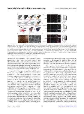
Figure 4. A study on a conduit made of a natural hydrogel double network supported by an aligned graphene mesh, loaded with Netrin-1, for peripheral
nerve regeneration. (A) Schematic image of netrin-1-loaded GMT/hydrogel nerve scaffold. (B) Optical image of the graphene mesh. (C) SEM image
of the graphene mesh. (D–F) The regenerative capacity of the peripheral nerve in groups of the autologous graft, scaffold, and scaffold + netrin-1.
(D) Immunofluorescent staining images of CD31 in the perineural suture and microvessel density by CD31 staining in the perineural suture.
(E) Immunofluorescent staining images of CD31 in the intraneural structure and microvessel density by CD31 staining in the intraneural structure.
(F) Immunofluorescence staining of S100 of sciatic nerves and S100 positive cells of sciatic nerves. Scale bars: B, D, E, F: 100 μm, C: 50 μm. The figures
were reproduced from Huang et al. . Copyright 2021, American Chemical Society.
[88]
GMT: Graphene mesh tube; SEM: Scanning electron microscopy.
measuring 50 μm in diameter. The in vitro study results repair with neuron differentiation and neurite elongation,
demonstrated that both PDA/RGD-SG/PCL and regardless of the presence of graphene. There was no
PDA/RGD-MG/PCL scaffolds facilitated attachment and significant difference between the expression of neural
proliferation of Schwann cells, as well as promoting axonal markers β-tubulin III and GFAP with or without graphene.
regrowth and remyelination following peripheral nerve rGO is generally considered less toxic than GO since
injury. In addition, the effectiveness of the repair process its surface has fewer oxygen-containing functional groups.
was found to increase with the loading of Schwann cells. GO is often synthesized through the oxidation of graphene,
In a study by Huang et al., graphene and GO-added which introduces carboxyl, epoxy, and hydroxyl groups onto
bioink were compared for their toxicity in nerve tissue the graphene surface. These groups can potentially induce
engineering [102] . The study examined the toxicity of GO toxicity by disrupting cell membranes and causing oxidative
compared to pluronic-modified graphene (G-P) on NSCs stress. rGO is produced by reducing GO, which removes
when mixed with polyurethane (PU). The results indicated some of these oxygen-containing functional groups,
that GO (25 μg/mL) was more toxic to NSCs than G-P, as making it less toxic than GO [103] . Vijayavenkataraman et
evidenced by a lower cell survival rate and viability. The al. developed PCL/rGO scaffolds using a biodegradable
lower oxygen consumption rate values in PU/GO 25 μg/mL hydrogel with excellent printability and long-term
were attributed to the disrupted sp structure of GO. The mechanical stability and can provide chemical and
2
gene expression levels of β-tubulin and GFAP for PU/GO biochemical signals to cells [104] . In this study, PC12 cells
and PU did not differ significantly, but there was a visible were used to evaluate the cell proliferation and neural
expression of β-tubulin and GFAP in protein expressions. differentiation potential of PCL and PCL/rGO scaffolds.
However, no expression was observed for NSCs in PU Results from the PrestoBlue assay indicated that PCL/rGO
alone, indicating that GO had a limited promotion effect scaffolds supported significantly higher cell proliferation
on the neural differentiation of NSCs. When NSCs were compared to pure PCL scaffolds due to the higher surface
encapsulated in hydrogels with or without graphene, no area to volume ratio of rGO. RT-PCR analysis revealed
apparent cytotoxicity was observed on a 2D surface, but that the PCL/rGO scaffolds upregulated the expression of
graphene slightly slowed cell growth. After 2 weeks of β3-tubulin, NF-H, and GAP-43 genes, which are crucial for
culturing, NSCs in hydrogels showed promising nerve neural differentiation. Furthermore, immunocytochemistry
Volume 2 Issue 2 (2023) 10 https://doi.org/10.36922/msam.0620