
Page 13 - MSAM-2-2
P. 13
Materials Science in Additive Manufacturing Union of 2D nanomaterials and 3D printing
A C D E
B
F H I J
G
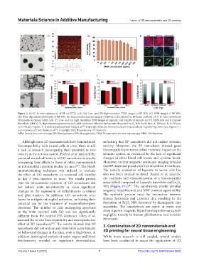
Figure 3. (A–E) In vitro cytotoxicity of BP on HT22 cells. (A) Low- and (B) high-resolution TEM images of BP NPs. (C) AFM images of BP NPs.
(D) Dose-dependent cytotoxicity of BP NPs. (E) Immunocytochemical analysis of HT22 cells cultured on BP-laden scaffolds. (F–J) In vitro cytotoxicity
of laponite on human GBM cells. (F) Low- and (G) high-resolution TEM images of laponite. Cell toxicity of laponite on (H) GBM cells and (I) human
fibroblasts (HFF1) (J) Hyperthermia generation and GBM antitumor effect of the laponite-decorated Fe O NPs. Scale bars: A: 200 μm, B, G: 50 μm,
4
3
[79]
E, F: 100 μm. Figures A–E were reproduced from Kang et al. . Copyright 2020, the Korean Society of Industrial and Engineering Chemistry. Figures F–J
were reproduced from Basina et al. . Copyright 2022, Royal Society of Chemistry.
[81]
AFM: Atomic force microscopy; BP: Blood pressure; NPs: Nanoparticles; TEM: Transmission electron microscopy; GBM: Glioblastoma.
Although some 2D nanomaterials have demonstrated indicating that BP nanosheets did not induce systemic
biocompatibility with neural cells in vitro, there is still toxicity. Moreover, the BP nanosheets showed good
a lack of research investigating their potential in vivo biocompatibility in terms of their minimal impact on the
toxicity in the nervous system. Portioli et al. analyzed the immune system, as evidenced by the lack of significant
potential neural cell toxicity of GO nanosheets in vivo by changes in white blood cell counts and cytokine levels.
comparing their effects to those of other nanomaterials Moreover, nuclear magnetic resonance imaging revealed
in intracerebral injection studies on mice . The NeuN that BP treatment posed a low risk of cerebral thrombosis.
[80]
immunostaining technique was utilized to evaluate The toxicity evaluation of laponite to nerve cells has
the effect of GO nanosheets on neuronal cell viability also not been studied in detail. Basina et al. describe
at day 2 post-injection in mice. The results proved the synthesis and characterization of a biocompatible
that the intracerebral injection of GO nanosheets did nano-hybrid composed of Laponite nanodisks and Fe O
4
3
[81]
not induce acute neurotoxicity or cause significant NPs (Figure 3F-J) . The nanohybrids exhibit ultrafast
changes in the expression of inflammatory cytokines magnetic hyperthermia and MRI contrast agent ability.
and glial markers. In addition, GO nanosheets were The synthetic process used the interaction between
found to mitigate microglial activation, indicating their ferrous hydroxide and Laponite clay, resulting in the
potential use for the treatment of neuroinflammatory formation of Fe O NPs decorated by diamagnetic clay
3
4
disorders. The number of cells undergoing apoptosis nanodisks. The nanohybrids are stable in water and
in the brain injected with GO was not significantly show superior magnetic hyperthermia performance with
different from the control (5% dextrose). Chen et al. negligible toxicity to human glioblastoma and foreskin
assessed the in vivo biocompatibility and neuroprotective fibroblasts.
effect of BP nanosheets . The results showed that BP 3. Combination of 2D nanomaterials and
[76]
nanosheets did not induce any observable acute toxicity
or behavioral changes in the mice, even at high doses. In 3D printing for neural tissue engineering
addition, histological analysis of major organs and blood While more research is still needed, several studies
biochemistry revealed no significant abnormalities, have been conducted to assess the application of 2D
Volume 2 Issue 2 (2023) 7 https://doi.org/10.36922/msam.0620