
Page 11 - MSAM-2-2
P. 11
Materials Science in Additive Manufacturing Union of 2D nanomaterials and 3D printing
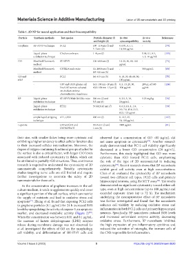
Table 1. 2D NP for neural application and their biocompatibility
Particle Synthesis methods Test species Particle diameter (l) In vitro In vivo References
and height (h) cytocompatibility toxicity
Graphene RF-cCVD technique PC12 100–110 nm (l) and 0, 0.01, 0.1, 1, - [73]
3–5 nm (h) 10,100 μg/mL
Liquid-phase Chicken embryos 1–4 μm (l) - 0.05, 0.1, 0.5, [115]
exfoliation technique 1, 5, 10 μg/mL
GO Modified Hummer’s SH-SY5Y 100–600 nm (l) 10, 20, 40, 80, 100 - [74]
method μg/mL
Modified Hummer’s C57BL/6 male mice 10–1800 nm (l) and - 500 μg/mL [80]
method 0.9–4.8 nm (h)
GO and - PC12 0.6–0.9 nm (h) 0, 10, 20, 40, 60, 80, - [75]
rGO 100 μg/mL
- U87 and U118 glioma cell GO: 100 nm–10 μm (l) 0, 5, 10, 20, 50, 200 μL of 500 [116]
line/U87 tumors cultured rGO: 100 nm–1.5 μm (l) 100 μg/mL μg/mL
on chicken embryo
chorioallantoic membrane
BP Liquid-phase SH-SY5Y/Male BALB/c mice 200 nm (l) and 0, 2.5, 5, 10, 5.25 mg/kg [76]
exfoliation technique 5.5 nm (h) 20 μg/mL
Liquid-phase HT22 57.60±6.43 nm (l) 0, 0.3, 0.5, 1, 1.9, - [79]
exfoliation technique 3.9, 7.8, 15.6, 31.3,
62.5, 125 μg/mL
simple liquid stripping 4T1, U251 200 nm (l) 0, 12.5, 25, - [117]
technique 50, 100 μg/mL
Laponite - U87wtEGFR and 30±5 nm (l) and 3000 μg/mL - [81]
U87EGFRvIII 1 nm (h)
their size, with smaller flakes being more cytotoxic and observed that a concentration of GO <80 μg/mL did
exhibiting a higher tendency to affect cellular function due not cause apoptosis or cytotoxicity . Another research
[74]
to their increased cellular internalization. Moreover, the study demonstrated that PC12 cell viability significantly
degree of oxygen-containing functional groups attached to decreased at a lower GO concentration (20 μg/mL).
the surface is also a critical factor, with larger C/O levels Furthermore, this study highlighted that GO was more
associated with reduced cytotoxicity in flakes, which can cytotoxic than rGO toward PC12 cells, emphasizing
be attributed to partially rGO structures. Thus, continuous the role of the type of 2D nanomaterial in inducing
research is required to understand the cytotoxicity of 2D cytotoxicity . Recent research shows that BP nanosheets
[75]
nanomaterials comprehensively. Notably, cytotoxicity exhibit good cell activity even at high concentrations.
studies targeting nerve cells are still limited and require Chen et al. evaluated the cytotoxicity of BP nanosheets
further investigations to ascertain the safety of 2D toward two different cell types, PC12 cells and primary
nanomaterials for these cells. hippocampal neurons, using the MTT assay . The results
[76]
As the concentration of graphene increases in the cell demonstrated no significant cytotoxicity toward either cell
culture medium, it tends to agglomerate quickly and cover type, even at high concentrations (up to 100 μg/mL) and
a significant portion of the cell surface, which can restrict extended exposure times (up to 72 h). The mechanism
the supply of nutrients and lead to oxidative stress and underlying the neuroprotective effect of BP nanosheets
apoptosis . Zhang et al. found that exposing PC12 cells was further investigated and found that the nanosheets
[72]
to graphene particles (0.1 μg/mL) for 24 h increased ROS enhance cell viability by reducing oxidative stress and
levels by upregulating the activity of caspase-3, an apoptotic inflammation in both PC12 cells and primary hippocampal
marker, and decreased metabolic activity (Figure 2) . neurons. Specifically, BP nanosheets reduced ROS levels
[73]
When the concentration was between 0.01 and 0.1 μg/mL, and increased antioxidant enzyme activity, decreasing
the increase of lactate dehydrogenase (LDH) enzyme oxidative stress. Furthermore, BP nanosheets inhibited
release during membrane damage was not observed. Lv the high expression of pro-inflammatory cytokines and
et al. investigated the effects of GO on the morphology, reduced the activation of microglia, the immune cells of
cell viability, and differentiation of SH-SY5Y cells and the CNS responsible for inflammation.
Volume 2 Issue 2 (2023) 5 https://doi.org/10.36922/msam.0620