
Page 14 - MSAM-2-2
P. 14
Materials Science in Additive Manufacturing Union of 2D nanomaterials and 3D printing
nanomaterials in bioink for nerve tissue regeneration critical role in nerve regeneration by promoting axonal
with 3D printing (Table 2). Zhu et al. synthesized hydrogel growth and guidance, as well as in the production
from gelatin methacrylamide and graphene nanoplatelets of ECM molecules and growth factors that support
(G-GelMA) [82] . Evaluating the cytotoxicity of the GelMA nerve regeneration [83,84] . This multipronged activity of
bioink incorporated with 1000 μg/mL of graphene, no Schwann cells makes them an attractive target for tissue
apparent cytotoxicity was observed compared to pure engineering research aimed at the regeneration and
[85]
GelMA bioink, but the cells proliferated with incubation repair of peripheral nerves . Uz et al. used 3D printing
time. Both hydrogels expressed abundant β-tubulin III to fabricate nerve regeneration conduits/scaffolds made
instead of GFAP, promoting differentiation of NSCs. of gelatin and graphene with tailored 3D microstructures
and mechanical properties . The study investigated how
[86]
Schwann cells have emerged as a promising research the microstructure of gelatin-based 3D scaffolds and
target for neural tissue engineering, particularly in nerve electrical stimuli affected mesenchymal stem cell (MSC)
regeneration. As a type of glial cell localized to the PNS, behavior and their transdifferentiation into Schwann cell-
Schwann cells are responsible for providing both physical like phenotypes. The gelatin-based 3D scaffolds exhibited
and metabolic support to neurons. Schwann cells play a favorable properties for MSC attachment and growth, and
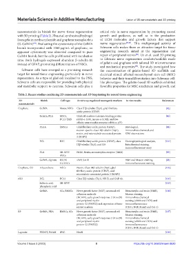
Table 2. Recent studies combining 2D nanomaterials and 3D bioprinting for neural tissue engineering
2D Bioink Cell type In vitro up-regulated neurogenic markers In vivo results References
nanomaterials
Graphene GelMA Mouse NSCs Class IIIβ-tubulin (Tuj1), glial fibrillary [82]
acidic protein (GFAP)
Gelatin, PLA MSCs, Glial cell markers (calcium-binding protein [86]
PC12-TrkB rabbit-α-S100, mouse-α-S100β, and low-
affinity neurotrophin receptor rabbit-α-p75)
PLGA hMSCs Glial fibrillary acidic protein (GFAP), Histological, [94]
neuron-specific class IIIβ-tubulin (Tuj1), immunohistochemical, and
nestin, and microtubule-associated protein SEM observations.
2 (MAP2)
PCL RSC Glial fibrillary acidic protein (GFAP), class Electrophysiological analysis, [101]
IIIβ-tubulin (Tuj1), and S10 histochemical staining,
immunofluorescent assay
PLA SH-SY5Y PAX6, Nestin, neurotrophin receptor (TrKB) [93]
iPSCs
GelMA, alginate RSC96 vWF, Sox10 H&E and Masson staining, [88]
HUVECs immunofluorescent staining
Graphene, GO Polyurethane NSCs Nestin, Class IIIβ-tubulin (Tuj1), glial [102]
fibrillary acidic protein (GFAP), and
microtubule-associated protein 2 (MAP2)
rGO PCL PC12 Class IIIβ-tubulin (Tuj1), NF-H, and GAP-43, [104]
Sulfuric acid, SH-SY5Y [105]
phosphoric acid
GelMA SCs, BMSCs Nerve growth factor (NGF), neuronal cell Hematoxylin and eosin (H&E), [106]
adhesion molecule Masson staining,
(NCAM), early growth response-2 (Krox20) immunohistochemical
and peripheral myelin staining (s100b and OCN) and
protein-22 (PMP22) and expression of bone- immunofluorescence
related markers (CD31, NGF, Runx2 and Col-1)
BP GelMA, PDA BMSCs, SCs Nerve growth factor (NGF), neuronal cell Hematoxylin and eosin (H&E), [107]
adhesion molecule Masson staining,
(NCAM), early growth response-2 (Krox20) immunohistochemical
and peripheral myelin staining (s100b and OCN) and
protein-22 (PMP22) immunofluorescence
(CD31, NGF, Runx2 and Col-1)
Laponite PEDOT, PAAM iPSC NeuN [109]
Volume 2 Issue 2 (2023) 8 https://doi.org/10.36922/msam.0620