
Page 60 - MSAM-2-2
P. 60
Materials Science in Additive Manufacturing Cellulose microfiber in ABS filament for 3D printing
3. Data and methods
This work presents considerations about a comparative
study between the mechanical properties of test specimens
formed by 3D-printing processing, using pure ABS and
ABS composite with highly crystalline cellulose additive.
3.1. Preparation of crystalline cellulose microfiber
The preparation of crystalline cellulose microfibers was
carried out from samples of E. grandis, certified by the
FSC, which were manually ground and dried at 110°C for
24 h in laboratory oven. The ground and dehydrated wood
was then subjected to acid hydrolysis, according to the
[36]
[35]
procedure by Qu et al. and Sanchez and Terence .
The samples were treated with nitric acid solution
(5 mol/L) for 300 min at 75°C (Figure 1). After the Figure 1. Acid hydrolysis of ground Eucalyptus grandis.
treatment with acid solution, the remaining material was
filtered under a 350-mmHg vacuum and washed with
deionized water until the pH of the filtered liquid was
neutral (Figure 2). A nylon #66 membrane was used as a
filtering element (0.2-µm porosity and 0.47-mm diameter)
due to its chemical resistance. A Hirch funnel was used
to support the filter membrane and facilitate the removal
of filtrate. The filtrates were dried in an oven for 24 h at
±60°C. The initial mass of ground and dehydrated wood
was 30.13 g, which allowed obtaining 15.38 g of cellulose
(51.04% yield).
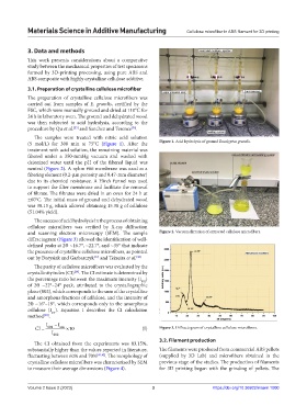
The success of acid hydrolysis in the process of obtaining
cellulose microfibers was verified by X-ray diffraction
and scanning electron microscopy (SEM). The sample Figure 2. Vacuum filtration of extracted cellulose microfibers.
diffractogram (Figure 3) allowed the identification of well-
defined peaks at 2ϴ ~16.7°, ~22.7°, and ~35° that indicate
the presence of crystalline cellulose microfibers, as pointed
out by Borysiak and Garbarczyk and Teixeira et al. [38]
[37]
The purity of cellulose microfibers was evaluated by the
crystallinity index (CI) . The CI estimate is determined by
[39]
the percentage ratio between the maximum intensity (I )
002
of 2ϴ ~22°–24° peak, attributed to the crystallographic
plane (002), which corresponds to the sum of the crystalline
and amorphous fractions of cellulose, and the intensity of
2ϴ ~16°–19°, which corresponds only to the amorphous
cellulose (I ). Equation I describes the CI calculation
am
[40]
method .
I I
CI 002 am 10 (I) Figure 3. Diffractogram of crystalline cellulose microfibers.
I 002
3.2. Filament production
The CI obtained from the experiments was 83.15%,
substantially higher than the values reported in literature, The filaments were produced from commercial ABS pellets
fluctuating between 60% and 70% [41,42] . The morphology of (supplied by 3D Lab) and microfibers obtained in the
crystalline cellulose microfibers was characterized by SEM previous stage of the studies. The production of filaments
to measure their average dimensions (Figure 4). for 3D printing began with the grinding of pellets. The
Volume 2 Issue 2 (2023) 3 https://doi.org/10.36922/msam.1000